
Page 696 - Williams Hematology ( PDFDrive )
P. 696
670 Part VI: The Erythrocyte Chapter 46: Erythrocyte Membrane Disorders 671
A B C D
E F F
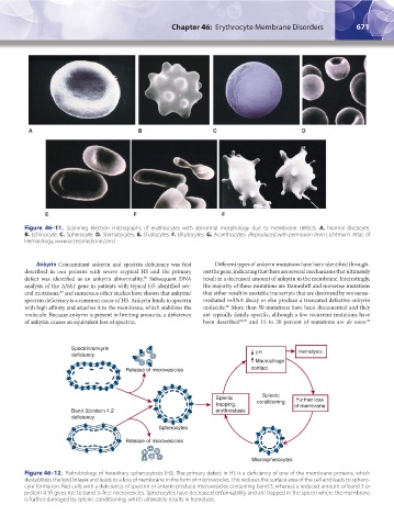
Figure 46–11. Scanning electron micrographs of erythrocytes with abnormal morphology due to membrane defects. A. Normal discocyte.
B. Echinocyte. C. Spherocyte. D. Stomatocytes. E. Ovalocytes. F. Elliptocytes. G. Acanthocytes. (Reproduced with permission from Lichtman’s Atlas of
Hematology, www.accessmedicine.com.)
Ankyrin Concomitant ankyrin and spectrin deficiency was first Different types of ankyrin mutations have been identified through-
described in two patients with severe atypical HS and the primary out the gene, indicating that there are several mechanisms that ultimately
defect was identified as an ankyrin abnormality. Subsequent DNA result in a decreased amount of ankyrin in the membrane. Interestingly,
68
analysis of the ANK1 gene in patients with typical HS identified sev- the majority of these mutations are frameshift and nonsense mutations
eral mutations, and numerous other studies have shown that ankyrin/ that either result in unstable transcripts that are destroyed by nonsense-
69
spectrin deficiency is a common cause of HS. Ankyrin binds to spectrin mediated mRNA decay or else produce a truncated defective ankyrin
with high affinity and attaches it to the membrane, which stabilizes the molecule. More than 50 mutations have been documented and they
66
molecule. Because ankyrin is present in limiting amounts, a deficiency are typically family-specific, although a few recurrent mutations have
of ankyrin causes an equivalent loss of spectrin. been described 69,70 and 15 to 20 percent of mutations are de novo.
63
Spectrin/ankyrin Hemolysis
deficiency pH
Macrophage
Release of microvesicles contact
Splenic
Splenic Further loss
trapping, conditioning of membrane
Band 3/protein 4.2 erythrostasis
deficiency
Spherocytes
Release of microvesicles
Microspherocytes
Figure 46–12. Pathobiology of hereditary spherocytosis (HS). The primary defect in HS is a deficiency of one of the membrane proteins, which
destabilizes the lipid bilayer and leads to a loss of membrane in the form of microvesicles. This reduces the surface area of the cell and leads to sphero-
cyte formation. Red cells with a deficiency of spectrin or ankyrin produce microvesicles containing band 3, whereas a reduced amount of band 3 or
protein 4.1R gives rise to band 3–free microvesicles. Spherocytes have decreased deformability and are trapped in the spleen where the membrane
is further damaged by splenic conditioning, which ultimately results in hemolysis.
Kaushansky_chapter 46_p0661-0688.indd 671 9/17/15 6:42 PM