
Page 693 - Williams Hematology ( PDFDrive )
P. 693
668 Part VI: The Erythrocyte Chapter 46: Erythrocyte Membrane Disorders 669
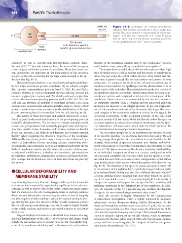
MARCKS Figure 46–8. Schematic of human erythrocyte
domain adducin. The domain structure for α and β adducin is
similar. The neck domain is responsible for oligomer-
39
N 9 kDa 30 kDa C ization and the tail represents the major binding
kDa
site for other red cell membrane proteins. MARCKS,
Tail domain myristoylated alanine-rich C kinase substrate.
actin filaments, spectrin,
Neck band 3, glucose transporter-1
domain
Head domain
tetramers, as well as tropomyosin, tropomodulin, adducin, dema- integrity of the membrane skeleton; and (3) the cytoplasmic viscosity,
tin, and p55. 19,21,50 Spectrin tetramers form the arms of the hexagons, which is determined primarily by intracellular haemoglobin. 54
cross-bridging individual junctional complexes. These horizontal pro- The unique biconcave disk shape of the erythrocyte provides a high
tein interactions are important in the maintenance of the structural ratio of surface area to cellular volume and this excess of membrane is
integrity of the cell, accounting for the high tensile strength of the ery- critical for survival of the cell. It enables the red cell to stretch and dis-
throcyte (see Fig. 46–1). tort when it passes through the microcirculation and protects it from
The spectrin/actin skeleton is anchored to the phospholipid bilayer destruction. To maintain the shape of the cell and to prevent loss of
by two major membrane protein complexes: (1) an ankyrin complex membrane microvesicles, the lipid bilayer and the skeleton have to be in
that contains transmembrane proteins, band 3, GPA, Rh, and RhAG direct contact with each other. The cohesion between the two sections of
complex proteins, as well as peripheral proteins ankyrin, protein 4.2, the membrane depends on protein–protein interactions between trans-
and several glycolytic enzymes, and (2) a distal junctional complex that membrane proteins and peripheral proteins in the vertical plane of the
contains the membrane-spanning proteins band 3, GPC, GLUT-1, Rh, membrane. These contacts are represented by the two macromolecu-
Kell, and XK proteins, in addition to peripheral proteins 4.1R, actin, lar complexes (ankyrin–band 3 complex and the junctional complex)
tropomyosin, tropomodulin, adducin, dematin, and p55. These vertical anchoring the skeleton to the integral proteins. To prevent fragmenta-
protein–protein interactions are critical in the stabilization of the lipid tion of the membrane and loss of the biconcave disk shape, the struc-
bilayer, preventing loss of microvesicles from the cells (see Fig. 46–1). tural integrity of the membrane skeleton is critical. In this regard, the
The avidity of these horizontal and vertical interactions is mod- horizontal interactions of the peripheral proteins of the junctional
ulated by posttranslational modifications of the participating proteins, complex, mainly 4.1R and actin, which link the tail ends of the spectrin
especially phosphorylation. The erythrocyte contains multiple protein tetramers together, is a major determinant of membrane stability. Spec-
kinases and phosphatases that constantly phosphorylate and dephos- trin heterodimer self-association, which links the head regions of the
phorylate specific serine, threonine, and tyrosine residues on band 3, spectrin tetramers, is also of paramount importance.
β-spectrin, ankyrin, 4.1R, adducin, and dematin, in a dynamic manner, The viscoelastic properties of the membrane are intrinsic features
thereby tightly regulating the structural properties of the membrane. of the spectrin skeleton. The enormous distortion imposed on the cell
Additionally, membrane protein associations are also influenced by a during passage through the microvasculature is accommodated by the
variety of intracellular factors, including calcium, calmodulin, phos- dynamic dissociation of spectrin tetramers into dimers, and subse-
phoinositides, and polyanions such as 2,3-bisphosphoglycerate (BPG). quent reassociation to restore the original shape once the shear stress is
Red cell membrane proteins are also subject to a variety of other post- removed. The lattice structure of the skeleton facilitates this flexibility,
55
translational modifications, including myristoylation, palmitoylation, as the individual hexagons are either in a compact configuration, with
glycosylation, methylation, deamidation, oxidation, and limited proteo- the junctional complexes close to each other and the spectrin tetram-
lytic cleavage, but the functional effects of these alterations are generally ers coiled between them, or in an extended configuration, which allows
not known. large unidirectional deformation without disruption of the skeleton (see
Fig. 46–4). The structure of the spectrin repeats also play a major role
CELLULAR DEFORMABILITY AND in the elasticity of the skeleton. Each triple helical repeat behaves partly
as an independently folding unit and has a different thermal stability.
56
MEMBRANE STABILITY Cysteine labeling studies indicated that shear stress forced the unfold-
ing of the least stable repeats. These studies highlight the flexibility of
57
In performing its primary function of oxygen delivery to the tissues, the the spectrin repeats and support the concept that their unfolding and
erythrocyte has to repeatedly negotiate tiny capillaries in the microvas- refolding contributes to the deformability of the membrane. In addi-
culature, as well as narrow slits in the spleen, which are much smaller tion, the elasticity of the ANK repeats may also facilitate the dynamic
than the diameter of the cell. Consequently, it has to undergo extensive changes in the membrane during circulatory shear stress. 22
distortion and deformation without fragmentation or loss of integrity, Red cell viscosity is largely determined by the concentration
and this property of deformability is critical for survival during its aver- of intracellular hemoglobin, which is tightly regulated to minimize
age 120-day life span. The structure of the red cell membrane endows cytoplasmic viscous dissipation during cellular deformation. As the
the cell with unique material properties, which makes it highly flexible, mean cell hemoglobin concentration rises above 37 g/dL, the viscosity
yet incredibly resilient, and enables a very rapid response to circulatory increases exponentially, and this compromises the deformability of the
shear stress. cell under increased circulatory shear stress. The hemoglobin concen-
54
Elegant biophysical studies have identified three features that reg- tration is critically dependent on red cell volume, which is primarily
ulate the deformability of the cell: (1) the biconcave disk shape, which determined by the total cation content of the cell. Numerous membrane
reflects the cell surface-area-to-volume ratio; (2) the viscoelastic prop- pumps and ion channels regulate the transport of sodium and potas-
erties of the membrane, which depend on the structural and functional sium across the membrane (Fig. 46–9).
Kaushansky_chapter 46_p0661-0688.indd 668 9/17/15 6:41 PM