
Page 769 - Williams Hematology ( PDFDrive )
P. 769
744 Part VI: The Erythrocyte Chapter 48: The Thalassemias: Disorders of Globin Synthesis 745
Osteoporosis is being recognized increasingly and may, at least in part,
be a reflection of hypogonadism. 201
β-THALASSEMIA INTERMEDIA
The clinical phenotype of patients designated as having thalassemia
intermedia is more severe than the usual asymptomatic thalassemia
trait but milder than transfusion-dependent thalassemia major. 7,199,200
The syndrome encompasses disorders with a wide spectrum of disabil-
ity. At the severe end, patients present with anemia later than patients
with the transfusion-dependent forms of homozygous β-thalassemia
and are just able to maintain a hemoglobin level of approximately 6 g/
dL without transfusion. However, their growth and development are
retarded. The patients become seriously disabled, with marked skeletal
deformities, arthritis, and bone pain; progressive splenomegaly; growth
retardation; and chronic ulcerations above the ankles. At the other end
of the spectrum, patients remain completely asymptomatic until adult
life and are transfusion independent, with hemoglobin levels as high as
10 to 12 g/dL. All varieties of intermediate severity are observed. Some
patients become disabled simply from the effects of hypersplenism.
Intensive studies of the molecular pathology of this condition have pro-
vided some guidelines about genotype–phenotype relationships that are
useful for genetic counseling (Table 48–6).
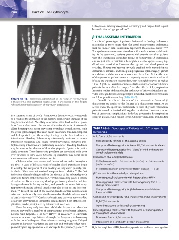
Figure 48–15. Radiologic appearances of the hands in homozygous Overall, the clinical features of the intermediate forms of β-
β-thalassemia. The scattered lucent areas in the bones of the fingers
reflect the marked expansion of marrow in distal areas. thalassemia are similar to the features of β-thalassemia major. At the
severe end of the spectrum, particularly in cases of growth retardation,
patients should be treated with regular transfusion. However, a num-
ber of important complications, including progressive hypersplenism,
is a common cause of death. Spontaneous fractures occur commonly occur in patients with milder forms. Clinically significant iron loading
as a result of the expansion of the marrow cavities with thinning of the
long bones and skull. Maxillary deformities often lead to dental prob-
lems from malocclusion. Formation of massive deposits of extramed-
ullary hematopoietic tissue may cause neurologic complications. With TABLE 48–6. Genotypes of Patients with β-Thalassemia
the gross splenomegaly that may occur, secondary thrombocytopenia Intermedia
and leukopenia frequently develop, leading to a further tendency to Mild forms of β-thalassemia
infection and bleeding. Splenectomy is frequently performed to reduce
+
transfusion frequency and severe thrombocytopenia; however, pos- Homozygosity for mild β -thalassemia alleles
7
tsplenectomy infections are particularly common. Bleeding tendency Compound heterozygosity for two mild β -thalassemia alleles
+
may be seen in the absence of thrombocytopenia. Epistaxis is partic- Compound heterozygosity for a “silent” or mild and more-se-
ularly common. These hemostatic problems are associated with poor vere β-thalassemia allele
liver function in some cases. Chronic leg ulceration may occur but is
more common in thalassemia intermedia. Inheritance of α- and β-thalassemia
Children who have grown and developed normally throughout β -Thalassemia with α -thalassemia (– –/αα) or α -thalassemia
+
0
+
the first 10 years of life as a result of regular blood transfusion begin (–α/αα or –α/–α)
to develop the symptoms of iron loading as they enter puberty, par- β -Thalassemia with genotype of Hgb H disease (– – /–α)
+
ticularly if they have not received adequate iron chelation. The first
7,9
indication of iron loading usually is the absence of the pubertal growth β-Thalassemia with elevated γ-chain synthesis
spurt and failure of the menarche. Over the succeeding years, a variety Homozygous β-thalassemia with heterocellular HPFH
of endocrine disturbances may develop, particularly diabetes mellitus, Homozygous β-thalassemia with homozygous γ 158 T→C
G
hypogonadotrophic hypogonadism, and growth hormone deficiency. change (some cases)
Hypothyroidism and adrenal insufficiency also occur but are less com-
mon. 7,186 Toward the end of the second decade, cardiac complications Compound heterozygosity for β-thalassemia and deletion
forms of HPFH
arise, and death usually occurs in the second or third decade as a result
of cardiac siderosis. 187–189 Cardiac siderosis may cause an acute cardiac Compound heterozygosity for β-thalassemia and β-chain variants
death with arrhythmia, or intractable cardiac failure. Both of these com- Hgb E/β-thalassemia
plications can be precipitated by intercurrent infection. Other interactions with rare β-chain variants
Even the adequately transfused child who has received chelation
therapy may suffer a number of complications. Bloodborne infection, Heterozygous β-thalassemia with triplicated or quadruplicated
notably with hepatitis B or C, HIV, or malaria, is extremely α-chain genes (ααα or αααα)
203
202
201
common in some populations, although the frequency is decreasing Dominant forms of β-thalassemia
with the use of widespread blood-donor screening programs. Delayed Interactions of β- and (δβ) - or (δβ) -thalassemia
0
+
puberty and growth retardation are common and probably reflect hypo-
gonadotrophic hypogonadism and damage to the pituitary gland. 201,204 Hgb, hemoglobin; HPFH, hereditary persistence of fetal hemoglobin.
Kaushansky_chapter 48_p0725-0758.indd 744 9/18/15 2:58 PM