
Page 803 - Williams Hematology ( PDFDrive )
P. 803
778 Part VI: The Erythrocyte Chapter 49: Disorders of Hemoglobin Structure: Sickle Cell Anemia and Related Abnormalities 779
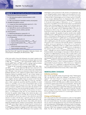
TABLE 49–5. Clinically Significant Hemoglobin Variants heterozygote is 40 to 45 percent. In the presence of α-thalassemia, neg-
atively charged β-globin variants compete more favorably for the avail-
I. Altered physical/chemical properties able α-chains; this phenomenon is reflected in even lower percentages
A. HbS (deoxyhemoglobin S polymerization): sickle of HbS and HbC in heterozygous carriers of these variants in the pres-
syndromes ence of common deletional forms of α-thalassemia (HbS of 30 to 35
+
B. HbC (crystallization): hemolytic anemia; microcytosis percent in individuals with heterozygous α -thalassemia, –α/αα; and 25
+
II. Unstable hemoglobin variants: to 30 percent in homozygous α -thalassemia, –α/–α). 396,397 Conversely,
A. Congenital Heinz body hemolytic anemia (N = 135) the amount of a β-globin variant will increase if there is a β-thalassemia
allele in trans; the percentage of the variant will be inversely propor-
III. Variants with altered oxygen affinity tional to the output of the β-thalassemia allele; thus, the higher the vari-
A. High-affinity variants: erythrocytosis (N = 92) ant the lower the output of the β -thalassemia allele. In the case of a
+
B. Low-affinity variants: anemia, cyanosis β -thalassemia allele in trans, the variant will amount to greater than
0
IV. M hemoglobins 90 percent or more of the Hb in red cells, with HbA and HbF consti-
2
A. Methemoglobinemia, cyanosis (N = 9) tuting the remainder. The quantity of α-globin variants is also variable,
V. Variants causing a thalassemic phenotype (N = 50) depending on the α-globin gene involved, and the presence of concom-
itant α- or β-thalassemia. Because there are normally four α-globin loci
A. β-Thalassemia (αα/αα) and the upstream 5′ α-globin gene (α ) is expressed at a higher
2
1. Hb (δβ) fusion (N = 3) level, some of the variation in the level of α-globin variants depends on
Lepore
2. Aberrant RNA processing (HbE, Hb , Hb ) which α-globin gene carries the mutation; α -globin mutations are usu-
Knossos Malay 2
3. Hyperunstable globins (Hb , Hb , etc.) ally present at 20 to 25 percent of the total Hb, whereas 3′ downstream
Geneva Westdale α -globin variants are expressed at a lower level (15 to 20 percent).
B. α-Thalassemia Concomitant α-thalassemia results in a higher level of expression of
1
1. Chain termination mutants (Hb )
Constant Spring α-globin variants. Observations on the different levels of expression of
2. Hyperunstable variants (Hb ) the common α-globin variant, HbG (α68Asn→Lys), is a case in
Quong Sze Philadelphia
point. Although this variant is found in approximately 25 percent of
398
Data from Bunn HF, Forget BG: Hemoglobin: Molecular, Genetic, and
Clinical Aspects. Philadelphia, PA: WB Saunders; 1986. Northern Italians, its percentages in Americans of African descent can
be either 33 or approximately 50 percent. This is clearly related to the
different genotypes found in these two distinct populations: In northern
Italy and Sardinia, the genotype is α α/αα, with an expression level of
G
of the fusion globin (hence the thalassemic phenotype), mutations that approximately 30 percent, whereas in Americans of African descent, the
cause both a missense mutation and create an aberrant splice site (such HbG Philadelphia mutation is commonly found on a hybrid α α gene asso-
2 1
+
G
as HbE, Hb Knossos , and Hb Malay ), and “hyperunstable” globins where the ciated with the common 3.7 kb α -thalassemia deletion (–α /αα) with
+
nascent globin chains are highly unstable, undergo rapid proteolytic approximately 33 percent expression. When there is an α -thalassemia
G
degradation, and result in a reduction in the affected globin. deletion in trans (–α /–α genotype), as expected, the level of HbG Philadel-
Except for the commonly occurring variants (HbS, HbC, HbE, phia will be approximately 50 percent. Coinheritance of α-chain variants
and HbD Los Angeles ), very few abnormal Hbs have been observed in the with β-thalassemia results in the increase of the α-chain variant.
homozygous state. Variant Hbs are usually found in the heterozygous
state. Although γ-chain variants are expressed in fetal life and their level HEMOGLOBIN C DISEASE
gradually decreases as the γ-globin to β-globin (fetal to adult) switch
progresses during the postnatal period, β- and α-chain variants are Definition and History
399
expressed throughout life. δ-Globin variants are expressed at very low HbC was the second Hb variant described after HbS. Homozygous
levels and can be detected only after the switch to adult globin synthe- HbC was described by Spaet and colleagues and Ranney and col-
400
sis is complete. Because α-globin chains are present in all of the Hbs leagues. HbC trait is found in 2 percent of Americans of African
401
expressed after the embryonic stage (HbF-α γ ; HbA-α β , and HbA - descent, and approximately one in 6000 have homozygous HbC.
402
2 2
2
2 2
α δ ), α-chain variants are associated with the production of variant Coinheritance of HbC with HbS results in HbSC disease, which is the
2 2
x
x
HbF (α γ ) and HbA (α δ ) as well. In heterozygous states, β-chain second most common form of SCD in the United States. There are also
2
2
2
2
2
variants constitute 40 to 50 percent of the Hb in red cells; it should, rare cases of HbC-β -thalassemia and HbC–β -thalassemia. HbC is
+
0
however, be kept in mind that certain factors affect the amount of vari- thought to have originated in Central West Africa and in parts of West
ant β chains in carriers. These factors include the stability of the variant, Africa, where the prevalence of HbC can reach 12.5 percent. The HbC
the surface charge of the variant β-chain, and the presence of concom- gene was found on three distinct β-globin cluster haplotypes, termed
itant α- or β-thalassemia (Chap. 48). The more unstable the variant, CI, CII, and CIII; the most common is CI, accounting for 70 percent or
the lower the quantity. Surface charge of the variant also plays a role more of HbC studied. 403
in determining the quantity in red cells; this is because the formation
of the αβ-dimers (α β and α β contacts) is the critical first step in Hb Etiology and Pathogenesis
2 2
1 1
tetramer formation, and this step is primarily driven by electrostatic HbC is the result of a GAG→AAG transition in codon 6 of the β-globin
interactions between α and β chains. The α-globin chains have a rela- gene, which changes the amino acid residue at this position from gluta-
tively positive surface charge, they interact more readily with relatively mic acid to lysine (Glu→Lys). The resultant positively charged Hb vari-
negatively charged β-globin variants to form αβ dimers. This is reflected ant can easily be distinguished from HbA and HbS by electrophoresis
in the higher percentage of negatively charged β-globin variants such as and chromatography, including high-performance liquid chromatogra-
HbN Baltimore (β95Lys→Glu), which is found in approximately 50 percent phy. HbC does not differ from HbA in its solubility; however, purified
in heterozygotes compared to β-globin variants with a positive surface solutions of HbC form tetragonal crystals in high-molarity phosphate
charge, HbS (β6Glu→Val) or HbC (β6Glu→Lys) whose quantity in the buffer. The Hb in red cells from homozygous HbC individuals also
Kaushansky_chapter 49_p0759-0788.indd 778 9/18/15 3:01 PM