
Page 804 - Williams Hematology ( PDFDrive )
P. 804
778 Part VI: The Erythrocyte Chapter 49: Disorders of Hemoglobin Structure: Sickle Cell Anemia and Related Abnormalities 779
A B C
D E F
G H I
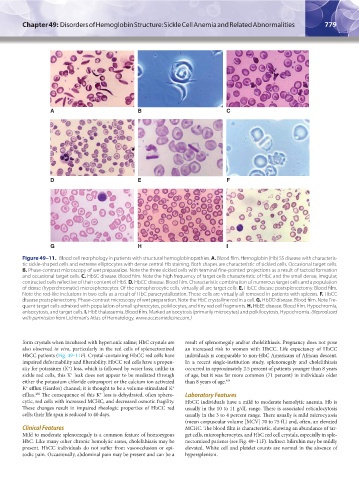
Figure 49–11. Blood cell morphology in patients with structural hemoglobinopathies. A. Blood film. Hemoglobin (Hb) SS disease with characteris-
tic sickle-shaped cells and extreme elliptocytes with dense central Hb staining. Both shapes are characteristic of sickled cells. Occasional target cells.
B. Phase-contrast microscopy of wet preparation. Note the three sickled cells with terminal fine-pointed projections as a result of tactoid formation
and occasional target cells. C. HbSC disease. Blood film. Note the high frequency of target cells characteristic of HbC and the small dense, irregular,
contracted cells reflective of their content of HbS. D. HbCC disease. Blood film. Characteristic combination of numerous target cells and a population
of dense (hyperchromatic) microspherocytes. Of the nonspherocytic cells, virtually all are target cells. E. HbCC disease postsplenectomy. Blood film.
Note the rod-like inclusions in two cells as a result of HbC paracrystallization. These cells are virtually all removed in patients with spleens. F. HbCC
disease postsplenectomy. Phase-contrast microscopy of wet preparation. Note the HbC crystalline rod in a cell. G. HbDD disease. Blood film. Note Fre-
quent target cells admixed with population of small spherocytes, poikilocytes, and tiny red cell fragments. H. HbEE disease. Blood film. Hypochromia,
anisocytosis, and target cells. I. HbE thalassemia. Blood film. Marked anisocytosis (primarily microcytes) and poikilocytosis. Hypochromia. (Reproduced
with permission from Lichtman’s Atlas of Hematology, www.accessmedicine.com.)
form crystals when incubated with hypertonic saline; HbC crystals are result of splenomegaly and/or cholelithiasis. Pregnancy does not pose
also observed in vivo, particularly in the red cells of splenectomized an increased risk to women with HbCC. Life expectancy of HbCC
HbCC patients (Fig. 49–11F). Crystal-containing HbCC red cells have individuals is comparable to non-HbC Americans of African descent.
impaired deformability and filterability. HbCC red cells have a propen- In a recent single-institution study, splenomegaly and cholelithiasis
+
sity for potassium (K ) loss, which is followed by water loss; unlike in occurred in approximately 2.5 percent of patients younger than 8 years
+
sickle red cells, this K leak does not appear to be mediated through of age, but it was far more common (71 percent) in individuals older
either the potassium chloride cotransport or the calcium ion activated than 8 years of age. 404
K efflux (Gardos) channel; it is thought to be a volume-stimulated K
+
+
402
+
efflux. The consequence of this K loss is dehydrated, often sphero- Laboratory Features
cytic, red cells with increased MCHC, and decreased osmotic fragility. HbCC individuals have a mild to moderate hemolytic anemia. Hb is
These changes result in impaired rheologic properties of HbCC red usually in the 10 to 11 g/dL range. There is associated reticulocytosis
cells; their life span is reduced to 40 days. usually in the 3 to 4 percent range. There usually is mild microcytosis
(mean corpuscular volume [MCV] 70 to 75 fL) and, often, an elevated
Clinical Features MCHC. The blood film is characteristic, showing an abundance of tar-
Mild to moderate splenomegaly is a common feature of homozygous get cells, microspherocytes, and HbC red cell crystals, especially in sple-
HbC. Like many other chronic hemolytic states, cholelithiasis may be nectomized patients (see Fig. 49–11F). Indirect bilirubin may be mildly
present. HbCC individuals do not suffer from vasoocclusion or epi- elevated. White cell and platelet counts are normal in the absence of
sodic pain. Occasionally, abdominal pain may be present and can be a hypersplenism.
Kaushansky_chapter 49_p0759-0788.indd 779 9/18/15 3:01 PM