
Page 901 - Williams Hematology ( PDFDrive )
P. 901
876 Part VI: The Erythrocyte Chapter 57: Primary and Secondary Erythrocytoses 877
Erythrocytosis of Eisenmenger Syndrome heme pocket are unstable and are associated with hemolytic anemia
Patients with right-to-left shunting (Eisenmenger syndrome) develop and cyanosis. Inheritance of these disorders is autosomal dominant.
a degree of erythrocytosis comparable to that observed with similar High-affinity hemoglobins result from mutations in any of three globin
degrees of desaturation at high altitudes. The hematologic changes genes; those from α-globin gene mutations are congenital and life-long.
85
associated with this syndrome include hyperviscosity caused by ery- β-Globin gene mutations are not apparent at birth but manifest after
throcytosis. Erythrocytosis is present in most patients, but excessive fetal to adult hemoglobin switching at approximately 6 months of life,
phlebotomy may cause microcytosis and some have attributed this while γ-globin gene mutations cause transient increase of hemoglobin
effect to the exacerbation of the symptoms of hyperviscosity. In view of concentration at birth lasting only about 6 months.
28
recent understanding of physiology of HIF regulation, it may not be the
microcytosis, per se, that is detrimental, but the induced iron deficiency Polycythemia Secondary to Red Cell Enzyme Deficiencies
that inhibits PHD2 and increases HIF, which then directly causes pul- Deficiencies of red cell enzymes in early steps of glycolysis sometimes
monary vasoconstriction and enhanced pulmonary vascular pressure cause a marked decrease in the levels of 2,3-BPG (Chap. 47). Occasion-
(Chaps. 32 and 34). ally, mild polycythemia occurs in patients with methemoglobinemia as
a result of cytochrome b reductase (methemoglobin reductase) defi-
5
Obstructive Sleep Apnea-Induced Syndrome ciency (Chap. 50).
In the colorfully named pickwickian syndrome, polycythemia is The same pathophysiology as that seen in high-affinity hemoglo-
86
characterized by its association with extreme obesity and somnolence. bins is also exhibited in mutations of the 2,3-BPG mutase gene, result-
Today, the more widely studied OSA may not always be associated with ing in low 2,3-BPG. Because these mutations are very rare, with only a
obesity but can, if severe, cause arterial hypoxemia, hypercapnia, som- single family comprehensively studied, it is not clear if the mode of
91
87
nolence, and secondary polycythemia. 88 inheritance is recessive or dominant.
This condition, as well as other high-affinity hemoglobins, can
Smoker’s Polycythemia only be conclusively confirmed by direct measurement of a hemoglo-
Heavy smoking will result in the formation of carboxyhemoglobin, bin dissociation curve, conveniently expressed as the partial pressure
which does not transport oxygen (Chap. 50), and causes an increase in of oxygen required to saturate 50 percent of hemoglobin (p50O ); when
2
oxygen affinity of the remaining normal hemoglobin. Carboxyhemo- equipment for this is not available, p50 can be estimated from pH, pO
2
globin increases in relationship to the number of cigarettes or cigars and hemoglobin oxygen saturation of venous blood. 92,93
smoked each day (Table 57–2). This leads to tissue hypoxia, erythropoi-
etin production, and stimulation of red cell production. Smoking may Chemically Induced Tissue Hypoxia
89
also cause a reduction in plasma volume. Either augmentation of red A number of chemicals have been suspected of causing histotoxic
90
cell mass or shrinkage of plasma volume could easily explain the rise in anoxia and secondary polycythemia, but the only chemical with a
the hematocrit. predictable capacity to cause erythrocytosis is cobalt. Cobalt admin-
83
istration increases erythropoietin production by increasing HIFs
Polycythemia Secondary to High-Affinity Hemoglobins (see Chap. 32). 94
Hemoglobins with certain amino acid substitutions manifest an
increased affinity for oxygen, producing tissue hypoxia and compen-
satory erythrocytosis (Chap. 49). Mutations affecting amino acids of INAPPROPRIATE POLYCYTHEMIAS
the α β -globin chain interface affect normal rotation within molecules Congenital Disorders of Hypoxia Sensing
1 2
and impair the rate of deoxygenation. Changes in the carboxyl terminal Chuvash Polycythemia Chuvash polycythemia is the only known
and penultimate amino acids also impair intramolecular motion and endemic congenital polycythemia. The condition is caused by an abnor-
tend to keep molecules in a high-affinity state. Alterations in the amino mality in the oxygen-sensing pathway and causes thrombotic and
acids lining the central cavity of hemoglobin destabilize the binding of hemorrhagic vascular complications that lead to early mortality; sur-
2,3-BPG in this cavity and lead to increased oxygen affinity (Chaps. 47 vival beyond age 65 years is uncommon. 48,95 Inheritance is autosomal
and 49). Finally, some heme pocket mutations interfere with deoxygen- recessive, and affected patients tend to have normal blood gases, nor-
ation. Most hemoglobins with a mutation involving amino acids in the mal calculated p50, normal to increased erythropoietin levels, absence
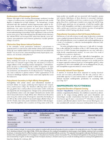
TABLE 57–2. Blood Oxygen Capacity in Smokers with Polycythemia
Hemoglobin (Hgb) Carboxyhemoglobin Affinity Correction
Subject (g/dL) (COHb) (g/dL) Hgb-COHb (g/dL) (g/dL) Adjusted Hgb (g/dL)
Healthy male 16 (14–18) 0.16 (0.08–0.25) 15.8 (14–18) 0 16 (14–18)
nonsmokers
Male smokers with 20 (17–23) 2 (1–3) 1816–21 1.5 (0.5–2.0) 16.5 (15–19)
increased Hgb
concentration
The male smokers include 10 consecutive subjects studied with elevated hematocrit and no evidence for polycythemia vera. The Hgb available
for O binding in blood is the Hgb-COHb. COHb also influences the residual hemoglobin to bind oxygen more tightly, making it less accessible
2
to tissues. A correction for this effect has been calculated and is labeled affinity correction. The adjusted hemoglobin indicates the blood concen-
trations that would be present in the subjects in the absence of excess carbon monoxide. Thus, the blood hemoglobin in this group of smokers
was increased by 3.5 g/dL on the average (from 16.5 [last column] to 20 [first column]) as a result of smoking-induced carboxyhemoglobinemia.
Data from Marshall A. Lichtman, MD.
Kaushansky_chapter 57_p0871-0888.indd 876 9/18/15 9:36 AM