
Page 903 - Williams Hematology ( PDFDrive )
P. 903
878 Part VI: The Erythrocyte Chapter 57: Primary and Secondary Erythrocytoses 879
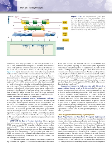
3p25 Figure 57–4. von Hippel-Lindau (VHL) gene
structure and mutation. Three exons of VHL genes
are depicted encoding for UTR (untranslated por-
EX1 EX2 EX3 DNA tion of mRNA), and coding sequences (CDs). VHL
domains β, α, β are shown. The relative number of
5 3 reported VHL gene mutations are depicted in verti-
UTR CDS UTR mRNA cal lines. The location of the Chuvash polycythemia
mutation is depicted by the diamond.
Amino
acid
N C
β α β Domain
Mutations
also develop acquired polycythemia. 99,111 The VHL gene codes for 213 It has been proposed that mutated VHL R200W protein hinders sup-
amino acids, and more than 130 germline mutations associated with pression of cytokine signaling SOCS1-mediated JAK2 degradation,
117
classic VHL syndrome have been identified, virtually all of them 5′ to via binding of a negative regulator of erythropoiesis, SOCS1, to the
112
the codon 200 position that is mutated in Chuvash polycythemia. extreme 3′ coding region of the VHL gene. Other observations are not
Figure 57–4 depicts the schematic effect of the Chuvash polycythemia consistent with this proposed mechanism: Another closely positioned
mutation in the context of other previously found VHL mutations. VHL polycythemia mutation, VHL H191D , is not associated with erythro-
It is not clear why mutations of a single gene lead to these two poietin hypersensitivity, while other, more upstream, mutations such
96
diverse phenotypes. It has been suggested that quantitative differences as VHL P138L are. Furthermore, the hypersensitivity of erythroid colo-
80
86
in loss of activity could explain the variable phenotypes among VHL nies is also seen in some HIF-2α mutations. Interestingly, in some, but
113
mutations, but the VHL gene may also have other functions, possibly not all, of these families, upregulation of NFE2, which enhances ery-
as a result of interactions with other modifying factors, that can contrib- thropoiesis, has been found. 6,118
ute to the onset of disease and that await future clarification. Another Unexplained Congenital Polycythemias with Elevated or
plausible explanation of polycythemia versus cancer predisposition Inappropriately Normal Levels of Erythropoietin The majority of
syndrome is that almost all polycythemic subjects have germline muta- patients with congenital polycythemias with inappropriately normal
tions of both VHL alleles, whereas those with VHL cancer predisposi- or elevated erythropoietin levels do not have VHL mutations, EGLN1
tion syndrome have only a single germline mutation and then acquire a or EPAS1 mutations, hemoglobinopathies, or 2,3-BPG deficiency,
somatic mutation that is essential for tumor genesis. and the molecular basis of polycythemia in these cases remains to be
EGLN1 Gene Mutations, Proline Hydroxylase Deficiency elucidated. Some such families show dominant inheritance, while
119
Another principal negative regulator of HIFs is PHD2 (encoded by the in others inheritance is recessive, and in some it is sporadic. Lesions
EGLN1 gene), which targets the α subunit of HIF for degradation. The in genes linked to hypoxia independent regulation of HIF, as well as
first loss-of-function mutation of PHD2 (PHD2 P317R ) was identified in a oxygen-dependent gene regulation pathways, are leading candidates for
family in which heterozygotes had mild or borderline polycythemia. mutation screening in polycythemic patients with normal or elevated
114
Since then, 25 additional patients with unexplained polycythemia erythropoietin without VHL, EGLN1 (proline hydroxylase), or EPAS1
who are heterozygote carriers of different PHD2 mutations have been (HIF-2α) mutations.
reported. Almost all patients with PHD2-associated polycythemia
115
have normal erythropoietin levels. Whether the cause of polycythemia Other Inappropriate Secondary Erythrocytoses
in this case is haploinsufficiency or a dominant negative effect remains Renal Polycythemia and Post–Renal Transplant Erythrocytosis
to be determined. Absolute erythrocytosis has been observed in a considerable number of
EPAS1 (HIF-2α) Gain-of-Function Mutations Affected patients patients with solitary renal cysts, polycystic renal disease, or hydrone-
have heterozygous missense mutations in the coding sequence of the phrosis. In most of these cases, erythropoietin assays on cyst fluid, serum,
EPAS1 gene that encodes HIF-2α, and typically have elevated erythro- or urine have disclosed the presence of erythropoietin. Patients with
120
poietin levels. 115,116 There is heterogeneity in these gain-of-function polycystic disease have a hematocrit value slightly higher than normal
HIF-2α mutations, but their existence supports the critical role of HIF- and definitely higher than would have been expected of patients with
2α in controlling the expression of erythropoietin. Some patients with uremia. In some patients on prolonged dialysis treatment, cystic trans-
EPAS1 mutations, similar to Chuvash polycythemia, have erythropoie- formation occurs in the native kidneys. This acquired cystic disease is
121
tin hypersensitive colonies, thus sharing features of both primary and occasionally associated with marked erythrocytosis. In patients with
secondary polycythemias. 97 pheochromocytoma or paraganglioma and erythrocytosis, erythropoie-
An explanation for the hypersensitivity of erythroid colonies bear- tin assays of serum and urine have disclosed higher-than-normal levels,
ing mutations that augment HIF stabilization remains to be discovered. and the erythrocytosis is most likely caused by excessive erythropoietin
Kaushansky_chapter 57_p0871-0888.indd 878 9/18/15 9:36 AM