
Page 951 - Williams Hematology ( PDFDrive )
P. 951
926 Part VII: Neutrophils, Eosinophils, Basophils, and Mast Cells Chapter 60: Structure and Composition of Neutrophils, Eosinophils, and Basophils 927
The violet-colored granules seen with light microscopy in mature
neutrophils on Wright-stained blood films are azurophilic granules
whose staining characteristics altered during maturation (Fig. 60–7). There-
fore, with light microscopy, the most reliable method for identifying
azurophilic granules on blood films is staining the cells for peroxidase.
Myeloblast The size of most of the peroxidase-negative granules (approximately 200
nm) is at the limit of resolution of the light microscope. The granules
cannot be distinguished individually but are responsible for the pink
background color of neutrophil cytoplasm during and after the myelo-
cyte stage.
Peroxidase-negative granules are more numerous than peroxidase-
positive granules during the myelocyte stage because peroxidase granule
Promyelocyte formation ceases after the promyelocyte stage, the number of oxidase-
positive granules per cell is reduced by mitoses, and peroxidase-negative gran-
ules continue to be produced by each myelocyte generation. 1
The purpose of nuclear segmentation is not known. Fluorescence
in situ hybridization with chromosome-specific probes has shown that
6
chromosomes are randomly distributed among the nuclear lobes. Some
mature neutrophils in women have drumstick- or club-shaped nuclear
Early myelocytes
appendages. These appendages contain the inactivated X chromosome.
An X-chromosome–specific nucleic acid probe has confirmed the posi-
tion of the X chromosomes in the drumstick structure of leukocyte
nuclei by in situ hybridization. 7
Late myelocytes
NEUTROPHIL GRANULES
The diversity of neutrophil granules appears to be linked to the timing
of biosynthesis during myelopoiesis. The hypothesis is that the different
subsets of granules are the result of differences in the biosynthetic win-
7
Metamyelocytes dows of the various granule proteins during maturation and not the
result of specific sorting between individual granule subsets (Chap. 66).
The control of biosynthesis is exerted by transcription factors that con-
trol the expression of the genes for the various granule proteins. Several
transcription factors identified as important in the timing of granule
protein synthesis, including the lineage-specific transcription factor
Band stage
GATA-1, the lineage-specific transcription factor PU.1, transcription
factor for various hematologic lineages, AML1 (also known as runt-re-
lated transcription factor 1 [RUNX1] or core-binding factor subunit
alpha-2 [CBFA2]), AML2 (also known as RUNX3), and AML3 (also
known as RUNX2 or CBFA1), and regulating factor of gene expres-
7–9
Neutrophil Eosinophil Basophil sion C/EBPε. The importance of C/EBPε has been emphasized by the
recognition of mutations in this protein in patients with the rare syn-
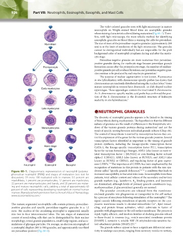
Figure 60–1. Diagrammatic representation of neutrophil (polymor- drome called “specific granule deficiency,” 10–12 a condition that leads to
phonuclear neutrophil [PMN]) and stages of maturation (see text for increased susceptibility to bacterial infections. In neutrophils from these
discussion). Of every 100 nucleated cells in marrow, 0.5 percent are patients, total cellular content and release of the secondary and tertiary
myeloblasts, 5 percent are promyelocytes, 12 percent are myelocytes, granule markers (e.g., lactoferrin, B binding protein, and lysozyme)
12
22 percent are metamyelocytes and bands, and 20 percent are matur- are diminished, although levels of primary granule constituents (e.g.,
ing and mature neutrophilic cells, yielding a total of approximately 60 myeloperoxidase, β-glucuronidase) generally are normal.
percent of cells representing developing neutrophils in normal human The granular constituents are released from the membrane-
marrow. (Reproduced with permission from Lichtman’s Atlas of Hematology. enclosed granules into phagosomes or transported to the cell surface
www.accessmedicine.com.)
by a process of exocytosis following stimulation of the neutrophil. The
13
signal cascade following stimulation of specific receptors on the cyto-
2+
The mature, segmented neutrophilic cells contain primary, peroxidase- plasmic membrane results in elevated intracellular Ca , lipid remod-
positive granules and specific peroxidase-negative granules in a 1:2 eling, and protein kinase activation, which culminate in fusion of
ratio. The nucleus of the circulating neutrophil is segmented, usually granules with phagosomes or the cell surface membrane. The process is
into two to four interconnected lobes. The late stages of maturation rapid, highly efficient, and involves families of docking proteins related
consist of nondividing cells that can be distinguished by their nuclear to those found in neurons (e.g., vesicle-associated membrane protein
morphology, mixed granule populations, small Golgi regions, and accu- [VAMP]-2, syntaxin-4, soluble NSF (N-ethylmaleimide-sensitive fac-
mulations of glycogen particles. On average, an electron micrograph of tor)-attachment protein [SNAP]-23). 14
a neutrophil displays 200 to 300 granules, and approximately one-third The granule subsets appear to have a significant differential sensi-
are peroxidase-positive (Fig. 60–6). tivity to undergo exocytosis, ranging from secretory vesicles to tertiary,
Kaushansky_chapter 60_p0923-0938.indd 926 9/18/15 10:34 PM