
Page 952 - Williams Hematology ( PDFDrive )
P. 952
926 Part VII: Neutrophils, Eosinophils, Basophils, and Mast Cells Chapter 60: Structure and Composition of Neutrophils, Eosinophils, and Basophils 927
A B
C D
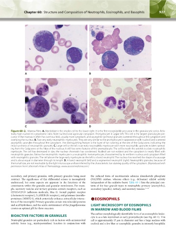
Figure 60–2. Marrow films. A. Myeloblast is the smaller cell to the lower right. It is the first recognizable precursor in the granulocytic series. Rela-
tively high nuclear-to-cytoplasmic ratio. Note nucleoli and agranular cytoplasm. Promyelocyte in upper left. This cell is the largest granulocyte pre-
cursor in the marrow. It often has overt nucleoli, usually more cytoplasm, and azurophilic (primary) granules scattered throughout the cytoplasm and
overlying the nucleus. B. Two very early neutrophilic myelocytes. They are very similar to the promyelocyte in appearance with nucleoli and scattered
azurophilic granules throughout the cytoplasm. The distinguishing feature is the burst of tan coloring at the site of the Golgi zone, indicating the
initial synthesis of neutrophilic granules. C. Large cell to the left is an early neutrophilic myelocyte with more neutrophilic granules evident spread-
ing from the Golgi zone at the hilus of the nucleus. It still has some features of the promyelocyte. The cell beneath the asterisk is a late neutrophilic
myelocyte. The cell has decreased in size, the nuclear chromatin has condensed. Nucleoli are not evident and the cytoplasm is nearly filled with
neutrophilic granules. Below the neutrophilic myelocyte is a neutrophilic metamyelocyte, characterized by its reniform nucleus and cytoplasm filled
with neutrophilic granules. The cell above the large early myelocyte on the left is a band neutrophil. The nucleus has reached the shape of a sausage
and is about equal in diameter through its length. D. A band neutrophil (left) and a segmented neutrophil (right). Neutrophilic granules, because of
their small size, are not resolvable by the light microscope and are inferred by the characteristic tan staining quality of the cytoplasm. (Reproduced with
permission from Lichtman’s Atlas of Hematology. www.accessmedicine.com.)
secondary, and primary granules, with primary granules being most the reduced form of nicotinamide adenine dinucleotide phosphate
resistant. The significance of this differential release is incompletely (NADPH) oxidase, whereas others (e.g., defensins) exhibit activity
understood, but some aspects are apparent in the functions of the independent of the oxidative burst. Table 60–1 lists the principal con-
constituents within the granules and granular membranes. For exam- tents of the four granule types in neutrophils: primary (azurophilic),
ple, secretory vesicles and tertiary granules contain receptors, such as secondary (specific), tertiary, and secretory vesicles. 15–56
CD11b/CD18 (adhesion molecule, Mac-1), formyl peptide receptor
(chemotactic receptor), FcγRIIIB (Fc receptor), and gelatinase (metallo-
proteinase [MMP]-9), which potentially enhance extracellular interac- EOSINOPHILS
tions of the neutrophil. Primary granules contain microbicidal proteins
and acid hydrolases, and the acidic environment of the phagolysosome LIGHT MICROSCOPY OF EOSINOPHILS
creates an optimal pH for these enzymes. IN MARROW AND BLOOD FILMS
BIOACTIVE FACTORS IN GRANULES The earliest morphologically identifiable form of an eosinophilic leuko-
cyte is as a late myeloblast or early promyelocyte (see Fig. 60–1). This
Neutrophil granules are particularly rich in factors with antimicrobial cell is approximately 15 µm in diameter and has a large nucleus with
activity. Some (e.g., myeloperoxidase) function in conjunction with nucleoli and a few blue or azurophilic granules in intensely basophilic
Kaushansky_chapter 60_p0923-0938.indd 927 9/18/15 10:34 PM