
Page 956 - Williams Hematology ( PDFDrive )
P. 956
930 Part VII: Neutrophils, Eosinophils, Basophils, and Mast Cells Chapter 60: Structure and Composition of Neutrophils, Eosinophils, and Basophils 931
Figure 60–8. Human mature eosinophil incubated for peroxidase.
Reaction product is present only in granules (g). The rough endoplas-
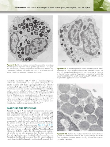
mic reticulum (er), including the perinuclear cisterna (pn) and the Golgi Figure 60–9. Mature basophil from human blood reacted for peroxi-
cisternae (Gc), does not contain reaction product. Most of the granules dase. Note the unusually large nucleus (N) and scattered glycogen par-
(arrow) contain the distinctive crystalline bar (×8000). ticles (gl). Human basophil granules contain peroxidase, as illustrated
by their density (as a result of the presence of reaction product) in this
type of preparation. They usually are spherical, difficult to fix, and may
be speckled in appearance (arrow) (×17,000).
bactericidal hypohalous acids. 66,67 ECP is a bactericidal protein
exists in two isoforms (ECP-1 and ECP-2) with activity toward hel-
minthic parasites. EDN shares high sequence homology with ECP
and is abundant, approximately 10 pg per cell. Other granule-stored
proteins include several enzymes of potential importance in inflam-
mation, including acid phosphatase, collagenase (MMP-8), matrix
metalloproteases, histaminase, catalase, and phospholipase D. 68–70
Chapter 62 discusses the functional aspects of these granular pro-
teins. In addition, mature eosinophils retain the ability to synthesize
a diverse array of proteins including cytokines and chemokines, 71,72
adhesion molecules, 73–76 receptors for cytokines, complement com-
ponents, lipid mediators, and immunoglobulins. 77–82
BASOPHILS AND MAST CELLS
Basophils (see Fig. 60–7) and mast cells were considered to be derived
from distinct lineages, but recent data indicates a common basophil-
mast cell progenitor exists from which mast cells exit the marrow as
immature precursors and terminally differentiate in tissues; basophils
mature in the marrow before entering the circulation. 83–86 This com-
mon progenitor is derived from the granulocyte–monocyte progenitor.
The granules of the two cell types stain metachromatically but are dis-
tinct when examined by electron microscopy (Figs. 60–9 and 60–10).
Identification of basophils in tissue at the light microscopic level is dif-
87
ficult without the use of cell specific antibodies. Basophils and mast
cells express the FcεR1 receptor. The cells can phagocytose sensitized
red cells but are less active phagocytes than the other granulocytes. Figure 60–10. Portion of a mast cell from human marrow. Note the
They lack significant amounts of antibacterial or lysosomal enzymes. granules are filled with scroll-like (s) and crystal (c) images and are dis-
Basophils are found in small numbers in blood (0.5 percent) and can be tinct from human basophil granules (see Fig. 60–9) in fine structural
seen in tissues in which inflammation resulting from hypersensitivity to morphology (×50,000).
Kaushansky_chapter 60_p0923-0938.indd 931 9/18/15 10:34 PM