
Page 954 - Williams Hematology ( PDFDrive )
P. 954
928 Part VII: Neutrophils, Eosinophils, Basophils, and Mast Cells Chapter 60: Structure and Composition of Neutrophils, Eosinophils, and Basophils 929
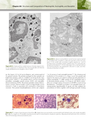
Figure 60–6. Mature neutrophil from normal human marrow reacted
for peroxidase. The cytoplasm is filled with granules of the two basic
types: (1) the smaller, pale, peroxidase-negative granules (p−) and (2)
the large, dense, peroxidase-positive granules (p+). The nucleus is con-
densed and lobulated (n –n ), the Golgi region (G) is small and without
4
1
Figure 60–5. Myelocyte from rabbit marrow in the late stage of mito- any forming granules, the endoplasmic reticulum is scant, and mito-
sis. This myelocyte is in telophase. Note that the granules are relatively chondria (m) are few (×21,000).
equally distributed to the daughter cells (×15,000).
are the largest, 0.5 to 0.8 µm in diameter, and contain much of 7 to 10 percent of total eosinophil protein. 62,63 The ultrastructural
the granular protein. The proteins packaged in these granules are localization of this protein is in a large, crystal-free granule and
highly basic proteins, with the crystalline core being mostly major supports the presence of a distinct primary granule population in
basic protein (MBP). 60,61 The granule matrix contains eosinophil mature eosinophils. 4,63,64 MBP consists of two homologues and is
peroxidase, eosinophil cationic protein (ECP), and eosinophil- an abundant granular protein, 5 to 10 pg per cell. Mature eosino-
derived neurotoxin (EDN). The primary granules contain Charcot- phils can no longer express this protein so all MBP is stored during
65
Leyden crystals. Charcot-Leyden crystals are bipyramidal crystals development. Eosinophil peroxidase is an abundant heme-con-
observed in fluids in association with eosinophilic inflammatory taining protein (approximately 15 pg per cell) that catalyzes the
reactions. They possess lysophospholipase activity and compose peroxidation of halides together with hydrogen peroxide forming
A B C
Figure 60–7. Images of granulocytes in blood films. A. Image shows two neutrophils, two eosinophils with bilobed nuclei, and a single neutrophil.
B and C. The images are of basophils showing densely stained metachromatic cytoplasmic granules. (Reproduced with permission from Lichtman’s
Atlas of Hematology. www.accessmedicine.com.)
Kaushansky_chapter 60_p0923-0938.indd 929 9/18/15 10:34 PM