
Page 953 - Williams Hematology ( PDFDrive )
P. 953
928 Part VII: Neutrophils, Eosinophils, Basophils, and Mast Cells Chapter 60: Structure and Composition of Neutrophils, Eosinophils, and Basophils 929
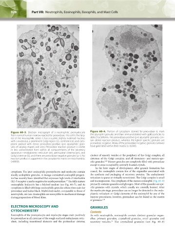
Figure 60–3. Electron micrograph of a neutrophilic promyelocyte Figure 60–4. Portion of cytoplasm stained for peroxidase to mark
from normal human marrow reacted for peroxidase. This cell is the larg- the azurophil granules and then immunolabeled with gold particles to
est of the neutrophilic series. It has a sizable, slightly indented nucleus detect lactoferrin. The peroxidase-positive (p+) azurophil granules con-
with a nucleolus, a prominent Golgi region (G), centriole (ce), and cyto- tain dense reaction product, whereas the lighter specific granules are
plasm packed with dense peroxidase-positive (p+) azurophilic gran- peroxidase negative. Many of the peroxidase-negative granules (arrows)
ules of varying shapes and sizes. Peroxidase reaction product is visible have gold label within their matrix (×70,000).
in less concentrated form within all compartments of the secretory
apparatus—endoplasmic reticulum (er), perinuclear cisterna (pn), and
Golgi cisternae (G), and there are peroxidase negative granules (p−). No clusters of smooth vesicles at the periphery of the Golgi complex; all
reaction product is apparent in the cytoplasmic matrix or mitochondria. cisternae of the Golgi complex; and all immature- and mature-spe-
(×8000). cific granules. Mature granules are completely filled with peroxidase
4,58
except in areas occupied by centrally located crystals.
In the later stages of development, after granule formation has
cytoplasm. The later eosinophilic promyelocyte and myelocyte contain ceased, the eosinophils contain few of the organelles associated with
mostly acidophilic granules. A lineage-committed eosinophil progeni- the synthesis and packaging of secretory proteins. The endoplasmic
tor has recently been identified that expresses high levels of interleukin reticulum is sparse or virtually nonexistent. The Golgi complex is small
(IL)-5 receptor α and is negative for myeloperoxidase. The fully mature and inconspicuous. The cytoplasm of the mature eosinophil (Fig. 60–8)
57
eosinophilic leukocyte has a bilobed nucleus (see Fig. 60–7), and its primarily contains granules and glycogen. Most of the granules are spe-
cytoplasm is filled with large eosinophilic granules whose rims stain for cific granules with crystals, which usually are centrally located. After
peroxidase and Sudan black. Multilobed nuclei, comparable to those of the myelocyte stage, peroxidase can no longer be detected in the endo-
neutrophils, are rare. Eosinophils are susceptible to mechanical damage plasmic reticulum or Golgi elements of the eosinophil by any of the
during preparation of blood films. enzyme procedures; however, peroxidase can be found in the matrix
of granules. 1,58
ELECTRON MICROSCOPY AND GRANULES
CYTOCHEMISTRY Contents
Eosinophils of the promyelocyte and myelocyte stages stain positively As with neutrophils, eosinophils contain distinct granular organ-
for peroxidase in all cisternae of the rough-surfaced endoplasmic retic- elles: primary granules, crystalloid granules, small granules and
ulum, including transitional elements and the perinuclear cisterna; secretory vesicles. The crystalloid granules (see Fig. 60–8)
59
Kaushansky_chapter 60_p0923-0938.indd 928 9/18/15 10:34 PM