
Page 99 - Williams Hematology ( PDFDrive )
P. 99
74 Part II: The Organization of the Lymphohematopoietic Tissues Chapter 5: Structure of the Marrow and the Hematopoietic Microenvironment 75
mitochondrial apoptosis pathway in the homeostasis of the hematopoi- without the involvement of the hematopoietic cytokines. HSC and their
etic cells populations in the marrow. 583,584 Antiapoptotic members of the myeloid and lymphoid progeny have multiple toll-like receptors (TLRs)
Bcl-2 family (Bcl-2, Bcl-X , Mcl-1, and A1) stabilize the mitochondrial which bind specific bacterial or viral molecules. 596,597 The activation of
L
membranes by preventing mitochondrial depolarization by the pore- TLRs leads to increased myelopoietic proliferation and differentiation,
585
forming family members, Bax and Bak. The antiapoptotic members especially of the monocyte/macrophage lineage, and differentiation
are also opposed by the proapoptotic, regulatory family members that of lymphoid cells toward the dendritic cell phenotype. 596,598 Although
consist of the BH3-only domain, such as Bim, Bid, Nix, and Puma. increased hematopoietic cytokines are produced by TLR activation, a
In HSC and multipotent progenitors, Mcl-1 is required to prevent direct response to TLR activation in hematopoietic cells changes the
apoptosis, and SCF stimulation increases the Mcl-1 expression. 586,587 prevalent myeloid transcription factor from C/EBPα, which mediates
In the later stages of single-lineage progenitors, Mcl-1 continues to be homeostasis by hematopoietic cytokines, to C/EBPβ, which mediates
required for survival of neutrophil and B and T lymphocytes, but it is the emergency responses to TLR activation. In response to the acti-
599
antagonized by the expression of Bim and Puma in these progenitors, vation of TLRs, mature neutrophils have decreased apoptosis as a result
providing a means to eliminate specific cells, such as autoreactive B and of increased Mcl-1 and decreased Bad activity. This may be a result
600
588
T lymphocytes. 583,584 A1 is required for normal neutrophil survival. of direct ligation of TLR receptors on LT-HSC, ST-HSC, and MPP that
In the erythroid lineage, Bcl-X is required to prevent apoptosis at are then stimulated to secrete cytokines such as IL-6, GM-CSF, and
L
the late erythroblast stage, and the proapoptotic Nix protein is also TNF-α. 601,602 An alternative path to apoptosis in hematopoietic cells is
589
590
expressed. The sequential proapoptotic and antiapoptotic stimuli the activation of specific death-domain receptors for the ligands such as
that regulate erythropoiesis demonstrate overlapping and cooperative FAS ligand, TNF-α, and TRAIL (tumor necrosis factor–related apopto-
interactions that affect erythroid cell homeostasis by both survival and sis-inducing ligand). Although these ligands are most commonly asso-
differentiation. Following moderate blood loss, an increased percent- ciated with pathologic states where they may play a role in the anemias
552
age of HSC enter cell cycle and self-renewal. In the BFU-E through of chronic disease, they have also been proposed to have a regulatory
CFU-E stages, SCF and glucocorticoids act in concert to upregulate role in normal erythropoiesis. 603
proliferation according to the erythropoietic requirements. How-
591
ever, because CFU-E depends upon EPO, SCF and EPO act together, REFERENCES
592
enhancing the proliferation and survival, respectively, of CFU-E. EPO
prevents apoptosis of CFU-E through basophilic erythroblast stages by 1. Neuman E: Ueber die Bedeutung des Knochenmarks für die Blutbildung. Cbl Med Wiss
decreasing Fas expression, 374,375 but its upregulation of Bcl-X prevents 6:689, 1868.
L
589
the apoptosis of the late-stage hemoglobin-producing erythroblasts. 2. Bizzozero G: Sulla funzione ematopoietica del midollo delle ossa. Gazz Med Ital Lomb
1:381, 1868.
Expression of proapoptotic Nix in very late erythroblasts and reticulo- 3. Neuman E: Du Role de la möelle des os dans la formation du sang. C R Acad Sci Paris
cytes plays a major role in targeting mitochondria for nontoxic elimina- 68:1112, 1869.
tion by autophagy. 593,594 4. Mosler F: Klinische Symptome und Therapie der medullären Leukämie. Berl Klin
Wochenschr 49:701, 1876.
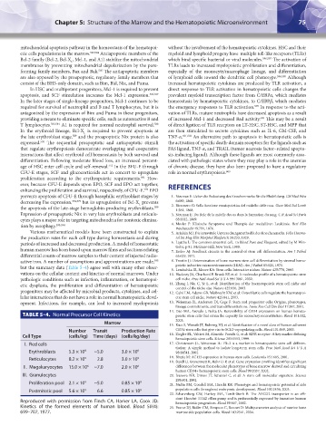
Various mathematical models have been constructed to explain 5. Arinkin MJ: Die intravitale Untersuchungsmethodik des Knochenmarks. Folia Haema-
the production rates for each cell type during homeostasis and during tol Int Mag Klin Morphol Blutforsch 38:233, 1929.
periods of increased and decreased production. A model of homeostatic 6. Lajatha L: The common ancestral cell, in Blood Pure and Eloquent, edited by M Win-
trobe, p 81. McGraw-Hill, New York, 1980.
human marrow has been based upon marrow films and sections relating 7. Erslev AJ: Feedback circuits in the control of stem cell differentiation. Am J Pathol
differential counts of marrow samples to their content of injected radio- 65:629, 1971.
active iron. A number of assumptions and approximations are made, 8. Trentin JJ: Determination of bone marrow stem cell differentiation by stromal hemo-
595
poietic inductive microenvironments (HIM). Am J Pathol 65:621, 1971.
but the summary data (Table 5–4) agree well with many other obser- 9. Lemischka IR, Moore KA: Stem cells: Interactive niches. Nature 425:778, 2003.
vations on the cellular content and kinetics of normal marrows. Under 10. Hackney JA, Charbord P, Brunk BP, et al: A molecular profile of a hematopoietic stem
pathologic conditions such as infection, inflammation, or hematopoi- cell niche. Proc Natl Acad Sci U S A 99:13061, 2002.
etic dysplasia, the proliferation and differentiation of hematopoietic 11. Zhang J, Niu C, Ye L, et al: Identification of the haematopoietic stem cell niche and
control of the niche size. Nature 425:836, 2003.
progenitors may be affected by microbial products, cytokines, and cel- 12. Calvi LM, Adams GB, Weibrecht KW, et al: Osteoblastic cells regulate the haematopoi-
lular interactions that do not have a role in normal hematopoietic devel- etic stem cell niche. Nature 425:841, 2003.
opment. Infections, for example, can lead to increased myelopoiesis 13. Weissman IL, Anderson DJ, Gage F: Stem and progenitor cells: Origins, phenotypes,
lineage commitments, and transdifferentiations. Annu Rev Cell Dev Biol 17:387, 2001.
14. Dao MA, Arevalo J, Nolta JA: Reversibility of CD34 expression on human hemato-
TABLE 5–4. Normal Precursor Cell Kinetics poietic stem cells that retain the capacity for secondary reconstitution. Blood 101:112,
2003.
Marrow 15. Kuci S, Wessels JT, Buhring HJ, et al: Identification of a novel class of human adherent
Number Transit Production Rate CD34-stem cells that give rise to SCID-repopulating cells. Blood 101:869, 2003.
Cell Type (cells/kg) Time (days) (cells/kg/day) 16. Ziegler BL, Valtieri M, Almeida-Porada G, et al: KDR receptor: A key marker defining
hematopoietic stem cells. Science 285:1553, 1999.
I. Red cells 17. Christensen JL, Weissman IL: Flk-2 is a marker in hematopoietic stem cell differen-
tiation: A simple method to isolate long-term stem cells. Proc Natl Acad Sci U S A
Erythroblasts 5.3 × 10 9 ~5.0 3.0 × 10 9 98:14541, 2001.
Reticulocytes 8.2 × 10 9 2.8 3.0 × 10 9 18. Bhatia M: AC133 expression in human stem cells. Leukemia 15:1685, 2001.
19. Steidl U, Kronenwett R, Rohr U-P, et al: Gene expression profiling identifies significant
II. Megakaryocytes 15.0 × 10 6 ~7.0 2.0 × 10 6 differences between the molecular phenotypes of bone marrow-derived and circulating
human CD34+ hematopoietic stem cells. Blood 99:2037, 2002.
III. Granulocytes 20. Ivanova NB, Dimos JT, Schaniel C, et al: A stem cell molecular signature. Science
298:601, 2002.
Proliferation pool 2.1 × 10 9 ~5.0 0.85 × 10 9 21. Nadin BM, Goodell MA, Hirschi KK: Phenotype and hematopoietic potential of side
Postmitotic pool 5.6 × 10 9 6.6 0.85 × 10 9 population cells throughout embryonic development. Blood 102:2436, 2003.
22. Scharenberg CW, Harkey MA, Torok-Storb B: The ABCG2 transporter is an effi-
cient Hoechst 33342 efflux pump and is preferentially expressed by immature human
Reproduced with permission from Finch CA, Harker LA, Cook JD: hematopoietic progenitors. Blood 99:507, 2002.
Kinetics of the formed elements of human blood. Blood 50(4): 23. Pearce DJ, Ridler CM, Simpson C, Bonnet D: Multiparameter analysis of murine bone
699–707, 1977. marrow side population cells. Blood 103:2541, 2004.
Kaushansky_chapter 05_p0051-0084.indd 75 9/19/15 12:11 AM