
Page 1025 - Clinical Immunology_ Principles and Practice ( PDFDrive )
P. 1025
988 Part seven Organ-Specific Inflammatory Disease
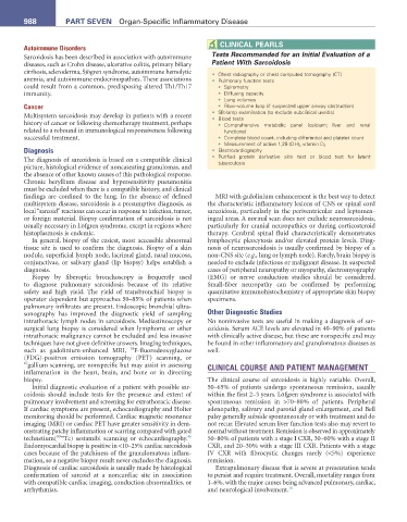
CLInICaL PearLs
Autoimmune Disorders
Sarcoidosis has been described in association with autoimmune Tests Recommended for an Initial Evaluation of a
diseases, such as Crohn disease, ulcerative colitis, primary biliary Patient With Sarcoidosis
cirrhosis, scleroderma, Sjögren syndrome, autoimmune hemolytic • Chest radiography or chest computed tomography (CT)
anemia, and autoimmune endocrinopathies. These associations • Pulmonary function tests
could result from a common, predisposing altered Th1/Th17 • Spirometry
immunity. • Diffusing capacity
• Lung volumes
Cancer • Flow–volume loop (if suspected upper airway obstruction)
• Slit-lamp examination (to exclude subclinical uveitis)
Multisystem sarcoidosis may develop in patients with a recent • Blood tests
history of cancer or following chemotherapy treatment, perhaps • Comprehensive metabolic panel (calcium; liver and renal
related to a rebound in immunological responsiveness following functions)
successful treatment. • Complete blood count, including differential and platelet count
• Measurement of active 1,25-(OH) 2 vitamin D 3
Diagnosis • Electrocardiography
The diagnosis of sarcoidosis is based on a compatible clinical • Purified protein derivative skin test or blood test for latent
tuberculosis
picture, histological evidence of noncaseating granulomas, and
the absence of other known causes of this pathological response.
Chronic beryllium disease and hypersensitivity pneumonitis
must be excluded when there is a compatible history, and clinical
findings are confined to the lung. In the absence of defined MRI with gadolinium enhancement is the best way to detect
multisystem disease, sarcoidosis is a presumptive diagnosis, as the characteristic inflammatory lesions of CNS or spinal cord
local “sarcoid” reactions can occur in response to infection, tumor, sarcoidosis, particularly in the periventricular and leptomen-
or foreign material. Biopsy confirmation of sarcoidosis is not ingeal areas. A normal scan does not exclude neurosarcoidosis,
usually necessary in Löfgren syndrome, except in regions where particularly for cranial neuropathies or during corticosteroid
histoplasmosis is endemic. therapy. Cerebral spinal fluid characteristically demonstrates
In general, biopsy of the easiest, most accessible abnormal lymphocytic pleocytosis and/or elevated protein levels. Diag-
tissue site is used to confirm the diagnosis. Biopsy of a skin nosis of neurosarcoidosis is usually confirmed by biopsy of a
nodule, superficial lymph node, lacrimal gland, nasal mucosa, non-CNS site (e.g., lung or lymph node). Rarely, brain biopsy is
conjunctivae, or salivary gland (lip biopsy) helps establish a needed to exclude infectious or malignant disease. In suspected
diagnosis. cases of peripheral neuropathy or myopathy, electromyography
Biopsy by fiberoptic bronchoscopy is frequently used (EMG) or nerve conduction studies should be considered.
to diagnose pulmonary sarcoidosis because of its relative Small-fiber neuropathy can be confirmed by performing
safety and high yield. The yield of transbronchial biopsy is quantitative immunohistochemistry of appropriate skin biopsy
operator dependent but approaches 50–85% of patients when specimens.
pulmonary infiltrates are present. Endoscopic bronchial ultra-
sonography has improved the diagnostic yield of sampling Other Diagnostic Studies
intrathoracic lymph nodes in sarcoidosis. Mediastinoscopy or No noninvasive tests are useful in making a diagnosis of sar-
surgical lung biopsy is considered when lymphoma or other coidosis. Serum ACE levels are elevated in 40–90% of patients
intrathoracic malignancy cannot be excluded and less invasive with clinically active disease, but these are nonspecific and may
techniques have not given definitive answers. Imaging techniques, be found in other inflammatory and granulomatous diseases as
18
such as gadolinium-enhanced MRI, F-fluorodeoxyglucose well.
(FDG)-positron emission tomography (PET) scanning, or
67 gallium scanning, are nonspecific but may assist in assessing CLINICAL COURSE AND PATIENT MANAGEMENT
inflammation in the heart, brain, and bone or in directing
biopsy. The clinical course of sarcoidosis is highly variable. Overall,
Initial diagnostic evaluation of a patient with possible sar- 50–65% of patients undergo spontaneous remission, usually
coidosis should include tests for the presence and extent of within the first 2–3 years. Löfgren syndrome is associated with
pulmonary involvement and screening for extrathoracic disease. spontaneous remission in >70–80% of patients. Peripheral
If cardiac symptoms are present, echocardiography and Holter adenopathy, salivary and parotid gland enlargement, and Bell
monitoring should be performed. Cardiac magnetic resonance palsy generally subside spontaneously or with treatment and do
imaging (MRI) or cardiac PET have greater sensitivity in dem- not recur. Elevated serum liver function tests also may revert to
onstrating patchy inflammation or scarring compared with gated normal without treatment. Remission is observed in approximately
46
technetium( 99m Tc) sestamibi scanning or echocardiography. 50–80% of patients with a stage I CXR, 30–60% with a stage II
Endomyocardial biopsy is positive in <10–25% cardiac sarcoidosis CXR, and 20–30% with a stage III CXR. Patients with a stage
cases because of the patchiness of the granulomatous inflam- IV CXR with fibrocystic changes rarely (<5%) experience
mation, so a negative biopsy result never excludes the diagnosis. remission.
Diagnosis of cardiac sarcoidosis is usually made by histological Extrapulmonary disease that is severe at presentation tends
confirmation of sarcoid at a noncardiac site in association to persist and require treatment. Overall, mortality ranges from
with compatible cardiac imaging, conduction abnormalities, or 1–6%, with the major causes being advanced pulmonary, cardiac,
arrhythmias. and neurological involvement. 10