
Page 1020 - Clinical Immunology_ Principles and Practice ( PDFDrive )
P. 1020
CHaPter 73 Sarcoidosis 983
IMMUNOLOGY/PATHOGENESIS anergy. Subsequently, bronchoalveolar lavage (BAL) studies in
pulmonary sarcoidosis showed that there were, in fact, enhanced
KeY COnCePts cell-mediated immune processes at sites of granulomatous
inflammation with activated CD4 BAL lymphocytes. These BAL
Immunopathogenesis of Sarcoidosis T cells are CD4 with a CD45RO “memory” phenotype expressing
+
+
high levels of activation molecules DR and VLA-1 (very late
• Noncaseating epithelioid granulomas
• Genetic susceptibility primarily involving major histocompatibility antigen-1, CD49a). Direct evidence that sarcoidosis is an antigen-
complex (MHC) genes driven disorder comes from studies of T-cell receptor (TCR)
• Oligoclonal expansion of T-cell receptor specific T cells consistent gene expression. These studies document the expansion of
with antigen-driven inflammation oligoclonal populations of T cells expressing specific α/β or γ/δ
• T-helper 1 (Th1) polarization at sites of disease TCR in BAL, blood, or skin (Kveim biopsy). The most compelling
• Decrease in regulatory T-cell (Treg) function contributes to Th1- data were derived from HLA-DRB1*03:01-positive Swedish
mediated inflammation +
• Potential role of interferon (IFN)-γ–expressing Th17.1 cells patients who have greatly increased numbers of AV2S3 Th1
+
• Potential role of classical Th17 effector cells cells in the lung. The expanded oligoclonal αβ T-cell subsets
• Etiology involves microbial triggers in sarcoidosis often contain shared amino acid motifs in the
• Mycobacterial or propionibacterial organisms most commonly CDR3 region of their Vβ or Vα/Jα genes, consistent with an
implicated antigen-driven T-cell response. The antigenic specificities of these
• Role of innate immunity in macrophage activation and granuloma expanded T-cell clones remain to be identified.
formation
• Progressive accumulation of serum amyloid A (SAA) within granulomas Th1 Polarization in Sarcoidosis
may drive chronic disease
• Nidus for granuloma formation T cells at sites of disease show a highly polarized Th1 cytokine
29
• Feed-forward amplification of local Th1 responses in part through profile at the time of diagnosis (Fig. 73.2). BAL studies have
Toll-like receptor 2 (TLR2) demonstrated expression of interferon-γ (IFN-γ) and tumor
necrosis factor (TNF) in pulmonary sarcoidosis. Consistent with
a polarized type 1 process, Th1-promoting cytokines interleukin-12
The histological hallmark of sarcoidosis is the presence of (IL-12), -18, -2, -15, and -27 are upregulated in sarcoidosis-affected
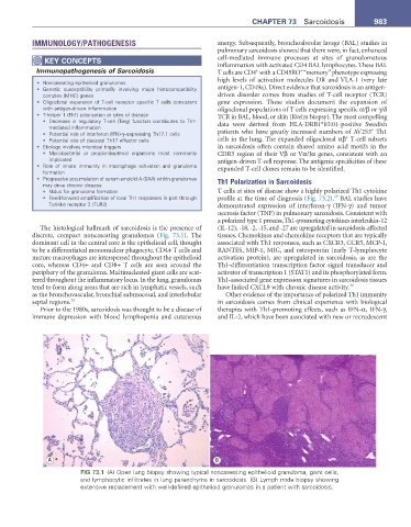
discrete, compact noncaseating granulomas (Fig. 73.1). The tissues. Chemokines and chemokine receptors that are typically
dominant cell in the central core is the epithelioid cell, thought associated with Th1 responses, such as CXCR3, CCR5, MCP-1,
to be a differentiated mononuclear phagocyte. CD4+ T cells and RANTES, MIP-1, MIG, and osteopontin (early T-lymphocyte
mature macrophages are interspersed throughout the epithelioid activation protein), are upregulated in sarcoidosis, as are the
core, whereas CD4+ and CD8+ T cells are seen around the Th1-differentiation transcription factor signal transducer and
periphery of the granuloma. Multinucleated giant cells are scat- activator of transcription 1 (STAT1) and its phosphorylated form.
tered throughout the inflammatory locus. In the lung, granulomas Th1-associated gene expression signatures in sarcoidosis tissues
tend to form along areas that are rich in lymphatic vessels, such have linked CXCL9 with chronic disease activity. 30
as the bronchovascular, bronchial submucosal, and interlobular Other evidence of the importance of polarized Th1 immunity
septal regions. 28 in sarcoidosis comes from clinical experience with biological
Prior to the 1980s, sarcoidosis was thought to be a disease of therapies with Th1-promoting effects, such as IFN-α, IFN-γ,
immune depression with blood lymphopenia and cutaneous and IL-2, which have been associated with new or recrudescent
A B
FIG 73.1 (A) Open lung biopsy showing typical noncaseating epithelioid granuloma, giant cells,
and lymphocytic infiltrates in lung parenchyma in sarcoidosis. (B) Lymph node biopsy showing
extensive replacement with well-defined epithelioid granulomas in a patient with sarcoidosis.