
Page 1038 - Clinical Immunology_ Principles and Practice ( PDFDrive )
P. 1038
CHaPter 74 Immunological Ocular Disease 1001
present clinically. The findings from one study, however, do The cornea is divided into five layers: the epithelium facing
support screening patients with scleritis with serological tests to the atmosphere, Bowman membrane, the stroma, Descemet
46
detect RA. Similarly, systemic lupus erythematosus (SLE) can membrane, and the endothelium abutting the aqueous humor.
be associated with scleritis, but one would not diagnose SLE on Opacification of the stroma is known as interstitial keratitis.
the basis of a positive ANA result, scleritis, and the absence of Congenital syphilis and other infections, such as herpes simplex,
other findings to suggest SLE. An erythrocyte sedimentation are important causes of the latter. Cogan syndrome is defined
rate (ESR) can sometimes be helpful in ensuring that a systemic as an autoimmune disease of the eighth nerve combined with
47
process is not present. Other laboratory tests are largely dictated interstitial keratitis, presumably on an autoimmune basis. Some
by the medication that is chosen to treat the scleritis. ophthalmologists also recognize autoimmune eighth-nerve disease
associated with other forms of ocular inflammation, such as
KERATITIS uveitis, as examples of Cogan syndrome. The pathogenesis of
Cogan syndrome is presumed to be a vasculitis, and many patients
The cornea is the anteriorly situated window to the eye. Normally, will have evidence for vasculitis elsewhere in the body. The
it should be clear. It can become opacified as a result of trauma, treatment usually requires aggressive use of systemic immunosup-
exposure to toxins, infection, dryness, calcium deposition, or pressive medications.
genetic diseases, such as corneal dystrophies. The three main
immune-mediated diseases that affect the cornea are peripheral CORNEAL TRANSPLANTATION AND
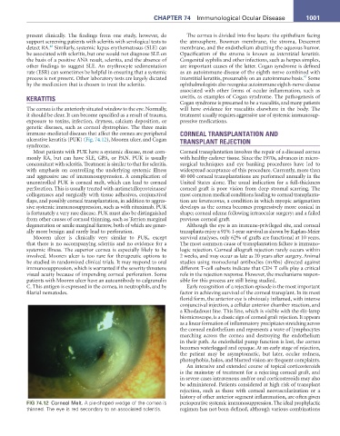
ulcerative keratitis (PUK) (Fig. 74.12), Mooren ulcer, and Cogan TRANSPLANT REJECTION
syndrome.
Most patients with PUK have a systemic disease, most com- Corneal transplantation involves the repair of a diseased cornea
monly RA, but can have SLE, GPA, or PAN. PUK is usually with healthy cadaver tissue. Since the 1970s, advances in micro-
concomitant with scleritis. Treatment is similar to that for scleritis, surgical techniques and eye banking procedures have led to
with emphasis on controlling the underlying systemic illness widespread acceptance of this procedure. Currently, more than
and aggressive use of immunosuppression. A complication of 40 000 corneal transplantations are performed annually in the
uncontrolled PUK is corneal melt, which can lead to corneal United States alone. The usual indication for a full-thickness
perforation. This is usually treated with antimetalloproteinases/ corneal graft is poor vision from deep stromal scarring. The
collagenases and surgically with tissue adhesives, conjunctival most common medical conditions leading to corneal transplanta-
flaps, and possibly corneal transplantation, in addition to aggres- tion are keratoconus, a condition in which myopic astigmatism
sive systemic immunosuppression, such as with rituximab. PUK develops as the cornea becomes progressively more conical in
is fortunately a very rare disease. PUK must also be distinguished shape; corneal edema following intraocular surgery; and a failed
from other causes of corneal thinning, such as Terrien marginal previous corneal graft.
degeneration or senile marginal furrow, both of which are gener- Although the eye is an immune-privileged site, and corneal
ally more benign and rarely lead to perforation. transplants enjoy a 91% 1-year survival as shown by Kaplan-Meier
Mooren ulcer is clinically very similar to PUK, except survival analyses, only 62% of grafts are functional at 10 years.
that there is no accompanying scleritis and no evidence for a The most common cause of transplantation failure is immuno-
systemic illness. The superior cornea is especially likely to be logic rejection. Corneal allograft rejection rarely occurs within
involved. Mooren ulcer is too rare for therapeutic options to 2 weeks, and may occur as late as 20 years after surgery. Animal
be studied in randomized clinical trials. It may respond to oral studies using monoclonal antibodies (mAbs) directed against
immunosuppression, which is warranted if the severity threatens different T-cell subsets indicate that CD4 T cells play a critical
visual acuity because of impending corneal perforation. Some role in the rejection response. However, the mechanisms respon-
patients with Mooren ulcer have an autoantibody to calgranulin sible for this process are still being studied.
C. This antigen is expressed in the cornea, in neutrophils, and by Early recognition of a rejection episode is the most important
filarial nematodes. factor in achieving survival of the corneal transplant. In its most
florid form, the anterior eye is obviously inflamed, with intense
conjunctival injection, a cellular anterior chamber reaction, and
a Khodadoust line. This line, which is visible with the slit-lamp
biomicroscope, is a classic sign of corneal graft rejection. It appears
as a linear formation of inflammatory precipitates stretching across
the corneal endothelium and represents a wave of lymphocytes
marching across the cornea and destroying the endothelium
in their path. As endothelial pump function is lost, the cornea
becomes waterlogged and opaque. At an early stage of rejection,
the patient may be asymptomatic, but later, ocular redness,
photophobia, halos, and blurred vision are frequent complaints.
An intensive and extended course of topical corticosteroids
is the mainstay of treatment for a rejecting corneal graft, and
in severe cases intravenous and/or oral corticosteroids may also
be administered. Patients considered at high risk of transplant
rejection, such as those with corneal neovascularization or a
history of other anterior segment inflammation, are often given
FIG 74.12 Corneal Melt. A pie-shaped wedge of the cornea is perioperative systemic immunosuppression. The ideal prophylactic
thinned. The eye is red secondary to an associated scleritis. regimen has not been defined, although various combinations