Page 1033 - Clinical Immunology_ Principles and Practice ( PDFDrive )
P. 1033

996          Part seven  Organ-Specific Inflammatory Disease


                                                                 TABLE 74.2  Characteristic Features of
                                                                 Common Forms of Uveitis
                                                                 Parameter
                                                                 Location   Anterior: ankylosing spondylitis, reactive arthritis,
                                                                             juvenile idiopathic arthritis
                                                                            Intermediate: Pars planitis
                                                                            Posterior: Vogt-Koyanagi-Harada syndrome (panuveitis
                                                                             most common)
                                                                 Onset      Sudden: ankylosing spondylitis, reactive arthritis
                                                                            Insidious: pars planitis, juvenile idiopathic arthritis
                                                                 Symmetry   Unilateral: ankylosing spondylitis, toxoplasmosis,
                                                                             herpetic
                                                                            Bilateral: pars planitis, lymphoma, juvenile idiopathic
                                                                             arthritis
                                                                 Course     Self-limiting: toxoplasmosis
                                                                            Recurrent: Behçet disease, ankylosing spondylitis
                                                                            Chronic: pars planitis
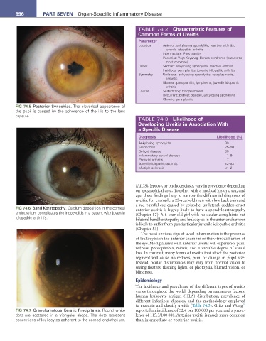
        FIG 74.5  Posterior Synechiae. The cloverleaf appearance of
        the pupil is caused by the adherence of the iris to the lens
        capsule.
                                                                 TABLE 74.3  Likelihood of
                                                                 Developing Uveitis in association With
                                                                 a specific Disease

                                                                 Diagnosis                           Likelihood (%)
                                                                 Ankylosing spondylitis                  30
                                                                 Sarcoidosis                             25–50
                                                                 Behçet disease                          80
                                                                 Inflammatory bowel disease              1–9
                                                                 Psoriatic arthritis                     7
                                                                 Juvenile idiopathic arthritis           <2–53
                                                                 Multiple sclerosis                      <1–2


                                                               (AIDS), leprosy, or onchocerciasis, vary in prevalence depending
                                                               on geographical area. Together with a medical history, sex, and
                                                               age, these findings help to narrow the differential diagnosis of
                                                               uveitis. For example, a 22-year-old man with low back pain and
                                                               a red painful eye caused by episodic, unilateral, sudden-onset
        FIG 74.6  Band Keratopathy. Calcium deposition in the corneal   anterior uveitis is highly likely to have a spondyloarthropathy
        endothelium complicates the iridocyclitis in a patient with juvenile   (Chapter 57). A 6-year-old girl with no ocular complaints but
        idiopathic arthritis.                                  bilateral band keratopathy and leukocytes in the anterior chamber
                                                               is likely to suffer from pauciarticular juvenile idiopathic arthritis
                                                               (Chapter 53).
                                                                  The most obvious sign of uveal inflammation is the presence
                                                               of leukocytes in the anterior chamber or the vitreous humor of
                                                               the eye. Most patients with anterior uveitis will experience pain,
                                                               redness, photophobia, miosis, and a variable degree of visual
                                                               loss. In contrast, many forms of uveitis that affect the posterior
                                                               segment will cause no redness, pain, or change in pupil size.
                                                               Instead, ocular disturbances may vary from normal vision to
                                                               seeing floaters, flashing lights, or photopsia, blurred vision, or
                                                               blindness.
                                                               Epidemiology
                                                               The incidence and prevalence of the different types of uveitis
                                                               varies throughout the world, depending on numerous factors:
                                                               human leukocyte antigen (HLA) distribution, prevalence of
                                                               different infectious diseases, and the methodology employed
                                                                                                                 14
                                                               to evaluate and classify uveitis (Table 74.3). Gritz and Wong
        FIG 74.7  Granulomatous Keratic Precipitates. Round white   reported an incidence of 52.4 per 100 000 per year and a preva-
        dots are scattered in a triangular shape. The dots represent   lence of 115.3/100 000. Anterior uveitis is much more common
        concretions of leukocytes adherent to the corneal endothelium.   than intermediate or posterior uveitis.
   1028   1029   1030   1031   1032   1033   1034   1035   1036   1037   1038