
Page 1035 - Clinical Immunology_ Principles and Practice ( PDFDrive )
P. 1035
998 Part seven Organ-Specific Inflammatory Disease
29
have arthritis ; and 7% of patients with psoriasis and arthritis
have uveitis. The diagnosis of psoriatic arthritis is made clinically
with the typical findings of cutaneous changes (erythematous,
hyperkeratotic rash) and joint disease. Patients with sacroiliac
disease and psoriatic arthritis are especially likely to develop
uveitis.
Sarcoidosis-Associated Uveitis
Ocular disease is the initial manifestation of sarcoidosis (Chapter
73) in 20% of patients. The systemic illness begins as ocular
inflammation almost as often as it initially presents as pulmonary
disease. Of the patients with sarcoidosis, 25–50% exhibit ocular
inflammation during the course of the illness. In most series,
sarcoidosis accounts for 3–10% of all patients with uveitis, second
to spondyloarthropathy as the most common systemic illness
associated with uveitis in North America and Europe. The
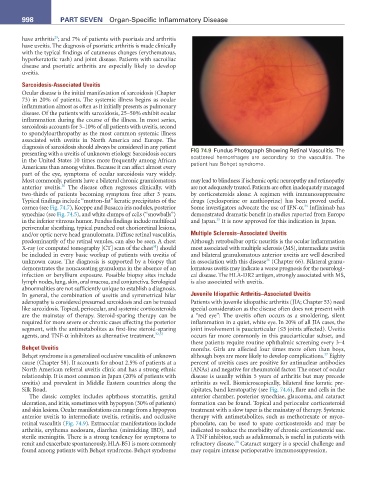
diagnosis of sarcoidosis should always be considered in any patient FIG 74.9 Fundus Photograph Showing Retinal Vasculitis. The
presenting with a uveitis of unknown etiology. Sarcoidosis occurs scattered hemorrhages are secondary to the vasculitis. The
in the United States 10 times more frequently among African patient has Behçet syndrome.
Americans than among whites. Because it can affect almost every
part of the eye, symptoms of ocular sarcoidosis vary widely.
Most commonly, patients have a bilateral chronic granulomatous may lead to blindness if ischemic optic neuropathy and retinopathy
30
anterior uveitis. The disease often regresses clinically, with are not adequately treated. Patients are often inadequately managed
two-thirds of patients becoming symptom free after 3 years. by corticosteroids alone: A regimen with immunosuppressive
Typical findings include “mutton-fat” keratic precipitates of the drugs (cyclosporine or azathioprine) has been proved useful.
34
cornea (see Fig. 74.7), Koeppe and Busacca iris nodules, posterior Some investigators advocate the use of IFN-α. Infliximab has
synechiae (see Fig. 74.5), and white clumps of cells (“snowballs”) demonstrated dramatic benefit in studies reported from Europe
35
in the inferior vitreous humor. Fundus findings include multifocal and Japan. It is now approved for this indication in Japan.
perivenular sheathing, typical punched out chorioretinal lesions,
and/or optic nerve head granulomata. Diffuse retinal vasculitis, Multiple Sclerosis–Associated Uveitis
predominantly of the retinal venules, can also be seen. A chest Although retrobulbar optic neuritis is the ocular inflammation
31
X-ray (or computed tomography [CT] scan of the chest ) should most associated with multiple sclerosis (MS), intermediate uveitis
be included in every basic workup of patients with uveitis of and bilateral granulomatous anterior uveitis are well described
36
unknown cause. The diagnosis is supported by a biopsy that in association with this disease (Chapter 66). Bilateral granu-
demonstrates the noncaseating granuloma in the absence of an lomatous uveitis may indicate a worse prognosis for the neurologi-
infection or beryllium exposure. Possible biopsy sites include cal disease. The HLA-DR2 antigen, strongly associated with MS,
lymph nodes, lung, skin, oral mucosa, and conjunctiva. Serological is also associated with uveitis.
abnormalities are not sufficiently unique to establish a diagnosis.
In general, the combination of uveitis and symmetrical hilar Juvenile Idiopathic Arthritis–Associated Uveitis
adenopathy is considered presumed sarcoidosis and can be treated Patients with juvenile idiopathic arthritis (JIA; Chapter 53) need
like sarcoidosis. Topical, periocular, and systemic corticosteroids special consideration as the disease often does not present with
are the mainstay of therapy. Steroid-sparing therapy can be a “red eye”: The uveitis often occurs as a smoldering, silent
required for more severe or chronic cases affecting the posterior inflammation in a quiet, white eye. In 20% of all JIA cases, the
segment, with the antimetabolites as first-line steroid-sparing joint involvement is pauciarticular (≤5 joints affected). Uveitis
agents, and TNF-α inhibitors as alternative treatment. 32,33 occurs far more commonly in this pauciarticular subset, and
these patients require routine ophthalmic screening every 3–4
Behçet Uveitis months. Girls are affected four times more often than boys,
37
Behçet syndrome is a generalized occlusive vasculitis of unknown although boys are more likely to develop complications. Eighty
cause (Chapter 58). It accounts for about 2.5% of patients at a percent of uveitis cases are positive for antinuclear antibodies
North American referral uveitis clinic and has a strong ethnic (ANAs) and negative for rheumatoid factor. The onset of ocular
relationship. It is most common in Japan (20% of patients with disease is usually within 5 years of arthritis but may precede
uveitis) and prevalent in Middle Eastern countries along the arthritis as well. Biomicroscopically, bilateral fine keratic pre-
Silk Road. cipitates, band keratopathy (see Fig. 74.6), flare and cells in the
The classic complex includes aphthous stomatitis, genital anterior chamber, posterior synechiae, glaucoma, and cataract
ulceration, and iritis, sometimes with hypopyon (30% of patients) formation can be found. Topical and periocular corticosteroid
and skin lesions. Ocular manifestations can range from a hypopyon treatment with a slow taper is the mainstay of therapy. Systemic
anterior uveitis to intermediate uveitis, retinitis, and occlusive therapy with antimetabolites, such as methotrexate or myco-
retinal vasculitis (Fig. 74.9). Extraocular manifestations include phenolate, can be used to spare corticosteroids and may be
arthritis, erythema nodosum, diarrhea (mimicking IBD), and indicated to reduce the morbidity of chronic corticosteroid use.
sterile meningitis. There is a strong tendency for symptoms to A TNF inhibitor, such as adalimumab, is useful in patients with
38
remit and exacerbate spontaneously. HLA-B51 is more commonly refractory disease. Cataract surgery is a special challenge and
found among patients with Behçet syndrome. Behçet syndrome may require intense perioperative immunosuppression.