
Page 1069 - Clinical Immunology_ Principles and Practice ( PDFDrive )
P. 1069
1034 ParT EighT Immunology of Neoplasia
Immune Other inhibitory
checkpoint therapy immune checkpoints
Anti-CTLA-4 TIM-3
Anti-PD-1/PD-L1 LAG-3
VISTA
Immune
Oncolytic virus costimulatory
immunotherapy molecules
ICOS
IMLYGIC (T-VEC)
4-1BB
OX40
Cancer Adoptive
vaccines cell therapy
Preventative vaccines Viral specific T cells
Therapeutic vaccines CAR T cells
T cells
Cytokine Monoclonal
therapy antibodies
IFN-α2b Conjugated mAbs
IL-2 Bispecific mAbs
Naked mAbs
Fig 77.1 Anticancer immunotherapeutic strategies.
(vii) Adoptive cell transfer, (viii) Monoclonal antibodies,
(ix) Cytokine therapy, (x) Cancer vaccines, (xi) Oncolytic virus
immunotherapy, (xii) Clinical challenges in immunotherapy,
and (xiii) Perspectives on future developments. Various anticancer
immunotherapeutic strategies are illustrated in Fig. 77.1.
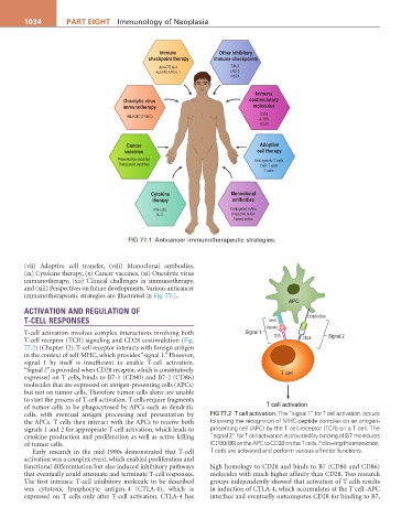
APC
ACTIVATION AND REGULATION OF
T-CELL RESPONSES MHC CD80/CD86
Peptide
T-cell activation involves complex interactions involving both Signal 1 TCR Signal 2
T-cell receptor (TCR) signaling and CD28 costimulation (Fig. CD28
77.2) (Chapter 12). T-cell receptor interacts with foreign antigen
in the context of self-MHC, which provides “signal 1.” However,
signal 1 by itself is insufficient to enable T-cell activation.
“Signal 2” is provided when CD28 receptor, which is constitutively T cell
expressed on T cells, binds to B7-1 (CD80) and B7-2 (CD86)
molecules that are expressed on antigen-presenting cells (APCs)
but not on tumor cells. Therefore tumor cells alone are unable
to start the process of T-cell activation. T cells require fragments
of tumor cells to be phagocytosed by APCs such as dendritic T cell activation
cells, with eventual antigen processing and presentation by Fig 77.2 T cell activation. The “signal 1” for T cell activation, occurs
the APCs. T cells then interact with the APCs to receive both following the recognition of MHC-peptide complex on an antigen-
signals 1 and 2 for appropriate T-cell activation, which leads to presenting cell (APC) by the T cell receptor (TCR) on a T cell. The
cytokine production and proliferation as well as active killing “signal 2” for T cell activation is provided by binding of B7 molecules
of tumor cells. (CD80/ 86) on the APC to CD28 on the T cells. Following this interaction,
Early research in the mid-1990s demonstrated that T-cell T cells are activated and perform various effector functions.
activation was a complex event, which enabled proliferation and
functional differentiation but also induced inhibitory pathways high homology to CD28 and binds to B7 (CD80 and CD86)
that eventually could attenuate and terminate T-cell responses. molecules with much higher affinity than CD28. Two research
The first intrinsic T-cell inhibitory molecule to be described groups independently showed that activation of T cells results
was cytotoxic lymphocyte antigen-4 (CTLA-4), which is in induction of CTLA-4, which accumulates at the T cell–APC
expressed on T cells only after T-cell activation. CTLA-4 has interface and eventually outcompetes CD28 for binding to B7,