
Page 1116 - Clinical Immunology_ Principles and Practice ( PDFDrive )
P. 1116
1080 Part eight Immunology of Neoplasia
MGUS, clonal lymphoplasmacytic cells must constitute <10% to idiopathic Bence-Jones proteinuria, light chain MM, AL
infiltration. amyloidosis, or LCDD.
Conventional cytogenetics are normal in patients with MGUS Patients with MGUS progress to a symptomatic plasma cell
due to technical limitations, a low proliferative rate, and the proliferative disorder or lymphoproliferative disorder at a rate
small number of plasma cells within the bone marrow samples. of 1% per year. However, not all persons with MGUS have the
However, studies using more sensitive methods have shown that same risk of disease progression. The risk of progression to a
chromosomal abnormalities are very common in the clonal cells more serious disease ranges widely from 0.6–3.4%/year based
found in MGUS. Analysis by interphase fluorescent in situ on the initial value of serum monoclonal protein as well as from
4
hybridization (FISH) demonstrate chromosomal aneuploidy, in 0.25–2.9%/year according to a risk stratification model. Three
particular hyperdiploidy, in approximately 50% of patients with adverse risk factors have been combined to create a risk stratifica-
MGUS. 1 tion model that is useful in predicting progression of MGUS
5
(non-IgM and IgM) to MM or a related malignancy : The findings
Diagnosis that generate a less favorable outcome include serum monoclonal
Diagnostic criteria for each of the individual types of MGUS protein level ≥1.5 g/dL, non-IgG MGUS, and abnormal serum
are determined by the International Myeloma Working Group free light chain ratio (kappa/lambda free light chain ratio <0.26
(IMWG) and are detailed in Table 80.1. 3 or >1.65).
The stratified absolute chance of disease progression over 20
Clinical Course years is 58% for high-risk MGUS associated with all three risk
The clinical course of MGUS depends largely on the specific factors, 37% for high- or intermediate-risk MGUS based on two
4
type. In non-IgM MGUS a minority of cases will progress to risk factors, 21% for low- or intermediate-risk MGUS associated
the more advanced premalignant stage SMM and to symptomatic with one risk factor, and 5% for low-risk MGUS based on no
5
MM. Less frequently, these patients progress to light chain risk factors. Risk factors for progression of LC-MGUS have yet
amyloidosis, light chain deposition disease (LCDD), or another to be defined.
lymphoproliferative disorder. IgM MGUS can progress to smolder-
ing WM (SWM) and to symptomatic WM. IgM MGUS also KeY CONCePtS
infrequently progresses to IgM MM. LC-MGUS may progress
MGUS
• A premalignant clonal plasma cell/lymphoplasmacytic proliferative
disorder.
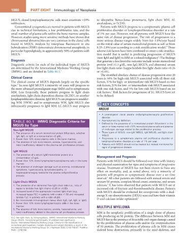
TABLE 80.1 iMWg Diagnostic Criteria for • Asymptomatic by definition.
MgUS by type • Defined by the presence of a monoclonal protein (M-protein) in the
serum or bone marrow with <10% monoclonal plasma cells and absence
Non-igM MgUS of end-organ damage related to the proliferative process.
1. The presence of a serum monoclonal protein (M-protein, whether • Three types of MGUS: non-IgM MGUS, IgM MGUS, and light chain
IgA, IgG, or IgD) at a concentration <3 g/dL. MGUS.
2. Fewer than 10% clonal plasma cells in the bone marrow. • Progresses to a symptomatic plasma cell proliferative disorder or
3. The absence of lytic bone lesions, anemia, hypercalcemia, and lymphoproliferative disorder at a rate of 1% per year.
renal insufficiency related to the plasma cell proliferative process. • Patients with MGUS should not be treated but instead monitored for
signs of progressive disease.
igM MgUS
1. The presence of a serum IgM monoclonal protein at a
concentration <3 g/dL. Management and Prognosis
2. Fewer than 10% clonal lymphoplasmacytic/plasma cells in the bone Patients with MGUS should be followed over time with history
marrow. and physical examination for signs and symptoms of progressive
3. The absence of end-organ damage such as anemia, constitutional disease. Treatment of MGUS has not been shown to have any
symptoms, hyperviscosity, lymphadenopathy, or
hepatosplenomegaly related to the plasma cell proliferative effect on mortality, and, as noted above, only a minority of
process. patients will progress to symptomatic disease over a set time
4
interval. All other patients are followed with annual serum and
Light Chain MgUS urinary M-protein, complete blood count, creatinine, and serum
4
1. The presence of an abnormal free light chain ratio (i.e., ratio of calcium. It has been observed that patients with MGUS are at
kappa to lambda free light chains <0.26 or >1.65). increased risk of fracture and thromboembolic disease. Patients
2. Increased level of the appropriate involved light chain (e.g., with MGUS should be evaluated for osteoporosis with a dual-
increased kappa FLC in patients with a ratio >1.65 and increased energy X-ray absorptiometry (DEXA) scan and have their vitamin
lambda FLC in patients with a ratio <0.26). 4
3. No monoclonal immunoglobulin heavy chain (IgG, IgA, IgD, or IgM). D and calcium intake optimized.
4. Fewer than 10% clonal lymphoplasmacytic cells in the bone
marrow. MULTIPLE MYELOMA
5. The absence of lytic bone lesions, anemia, hypercalcemia, and
renal insufficiency related to the plasma cell proliferative process. MM is the neoplastic proliferation of a single clone of plasma
cells producing an M-protein. The difference between MM and
FLC, free light chain; Ig, immunoglobulin; IMWG, International Myeloma Working
Group; MGUS, monoclonal gammopathy of undetermined significance. MGUS lies in the presence of detectable end-organ damage caused
Source: International Myeloma Working Group. Criteria for the classification of by the proliferation of plasma cells and/or the overproduction
monoclonal gammopathies, multiple myeloma and related disorders: a report of the of M-protein. The proliferation of plasma cells in MM causes
International Myeloma Working Group. Br J Haematol 2003;121(5):749–57. PubMed
PMID: 12780789. skeletal bone destruction, primarily in the axial skeleton, and