
Page 1118 - Clinical Immunology_ Principles and Practice ( PDFDrive )
P. 1118
1082 Part eight Immunology of Neoplasia
Ref G A M K L
0.305
0.290
0.274
0.259
0.244
0.229
0.214
0.198
0.183
0.168
0.153
0.137
0.122
0.107
0.091
0.078
0.061
0.048
0.030
0.015
0.000 B
A 0 20 40 60 80 100 120 140 160 180 200 220 240 260 280 300
Ref G A M K L
0.40
0.38 1
0.36
0.34
0.32
0.30
0.28
0.26
0.24
0.22
0.20
0.18
0.16
0.14
0.12
0.10
0.08
0.06
0.04
0.02
0.00
0 20 40 60 80 100 120 140 160 180 200 220 240 260 280 300
C D
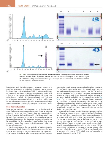
Fig 80.1 Electropherogram (A) and Immunofixation Electrophoresis (B) of Serum from a
Normal Patient and a Myeloma Patient (C) and (D). Note the M-spike in the gamma region
of the electropherogram and the immunoglobulin G (IgG) kappa band visible in the immunofixation
of the myeloma patient specimen.
leukopenia, and thrombocytopenia. Rouleaux formation is Mature plasma cells are oval with abundant basophilic cytoplasm.
particularly common in patients with elevated serum protein The nucleus is round and eccentrically located with a marked
levels, as noted in the discussion of MGUS. Monoclonal plasma perinuclear cytoplasmic clearing, termed a hof. 80. The nucleus
cells are rarely seen in the peripheral smear in patients with MM; contains “clock face“ or “spoke wheel“ chromatin without nucleoli.
a detectable absolute peripheral blood plasma cell count ≥100 Immature plasma cells have dispersed nuclear chromatin,
cells/µL (≥0.1 × 10/L) is found in approximately 10%. Evaluation prominent nucleoli, and a high nuclear to cytoplasmic ratio (Fig.
6
for circulating monoclonal plasma cells utilizes a slide-based 80.3). The cytoplasm of myeloma cells may contain condensed
immunofluorescence assay, a two-color immunoassay technique or crystallized cytoplasmic immunoglobulin resulting in the
+
−
(ELISPOT), or flow cytometry by gating on CD38 /CD45 cells. 6 following unusual findings, which are not limited to MM: multiple
pale bluish-white, grapelike accumulations, cherry-red refractive
Bone Marrow Evaluation round bodies, vermilion staining glycogen-rich IgA, overstuffed
Bone marrow aspirates and biopsies are key to the diagnosis of fibrils, and crystalline rods.
MM. The bone marrow plasma cell percentage should be estimated Immunohistochemical staining, immunofluorescent studies,
from a core biopsy specimen. However, if the percentage of plasma and flow cytometry detect either kappa or lambda light chains,
cells in the aspirate and core biopsy differ, the higher value should but not both, in the cytoplasm of bone marrow plasma cells
be used. Flow cytometry is not used to determine bone marrow in patients with MM while surface immunoglobulin is absent.
plasma cell percentage for diagnostic purposes. Clonality can The normal kappa/lambda ratio in the bone marrow is 2 : 1. A
be established by demonstrating kappa/lambda light chain ratio of more than 4 : 1 or less than 1 : 2 is considered to meet
6
restriction using flow cytometry, immunohistochemistry, or the definition of kappa or lambda monoclonality, respectively.
immunofluorescence. 6 As with normal plasma cells, myeloma cells express CD79a,
The bone marrow of the vast majority of patients contains p63, CD138, and CD38. In contrast to normal plasma cells,
10% or more clonal plasma cells. However, due to patchy bone myeloma cells infrequently express CD19. Approximately 70%
marrow involvement, a bone marrow aspirate and biopsy may of myeloma cells express CD56, which is typically negative in
6
show less than 10% plasma cells in approximately 4% of patients. normal plasma cells and in PCL. 6