
Page 1121 - Clinical Immunology_ Principles and Practice ( PDFDrive )
P. 1121
ChaPter 80 Monoclonal Gammopathies 1085
A B
C
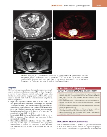
Fig 80.5 Myeloma lytic bone lesions visible in the sacral vertebra by (A) conventional computed
tomography (CT), (B) positron emission tomography (PET)/CT scans, and (C) magnetic resonance
imaging (MRI) demonstrate hypermetabolism in the sacrum. (Courtesy Dr. Jonathan Landa,
Department of Radiology, Memorial Sloan Kettering Cancer Center.)
Prognosis theraPeUtiC PriNCiPLeS
MM is a heterogeneous disease. Some patients progress rapidly Current Treatment of Multiple Myeloma (MM)
despite treatment, and others remain stable without therapy for
a number of years. Myeloma patients are typically staged using • Patients with standard- or intermediate-risk myeloma: High-dose
the Revised International Staging System (R-ISS). This system chemotherapy with autologous hematopoietic stem cell transplantation
stratifies patients with MM into three main risk categories at (HSCT) with induction chemotherapy regimen dependent on risk
time of diagnosis. 10 stratification.
• High-Risk Myeloma: Patients with t(14;16), t(14;20), or • Patients ineligible for HSCT: induction lenalidomide plus dexamethasone.
• Patients with high-risk MM do poorly with all conventional treatment
del17p13 by FISH are considered to have high-risk myeloma. options.
These patients have a median survival of approximately 2–3 • Patients should be evaluated before each treatment cycle to determine
years despite standard treatment. Patients with lactate dehy- degree of disease response.
drogenase (LDH) ≥2 times the institutional laboratory upper • Patients with one or more lesions on skeletal radiographs and those
limit of normal and those with features of primary are also with osteopenia should be given bisphosphonate therapy.
considered to have high-risk MM. • Almost all patients with MM who survive initial treatment will eventually
relapse and require further therapy.
• Intermediate-Risk Myeloma: Patients with t(4;14) or 1q+ by
FISH or deletion 13/hypodiploidy are considered to have
intermediate-risk MM.
• Standard-Risk Myeloma: All other patients with MM who SMOLDERING MULTIPLE MYELOMA
lack any of the high- or intermediate-risk cytogenetic
abnormalities or features are considered to have standard- SMM is defined as follows: M-protein ≥3 g/dL in serum and/or
risk MM; this includes patients with trisomies, t(11;14) and 10–60% bone marrow plasma cells, no end-organ damage (lytic
t(6;14). lesions, anemia, renal disease, or hypercalcemia) attributable to