
Page 1119 - Clinical Immunology_ Principles and Practice ( PDFDrive )
P. 1119
ChaPter 80 Monoclonal Gammopathies 1083
Ref GAM K L fK fL
0.200
0.190
0.180
0.170
0.160
0.150
0.140
0.130
0.120
0.110
0.100
0.090
0.080
0.070
0.060
0.050
0.040
0.030
0.020
0.010
0.000 B
A 0 20 40 60 80 100 120 140 160 180 200 220 240 260 280 300
0.200 Ref GAM K L fK fL
0.190
0.180
0.170
0.160
0.150
0.140
0.130
0.120
0.110
0.100
0.090
0.080
0.070
0.060
0.050
0.040 1
0.030
0.020
0.010
0.000
D
0 20 40 60 80 100 120 140 160 180 200 220 240 260 280 300
C
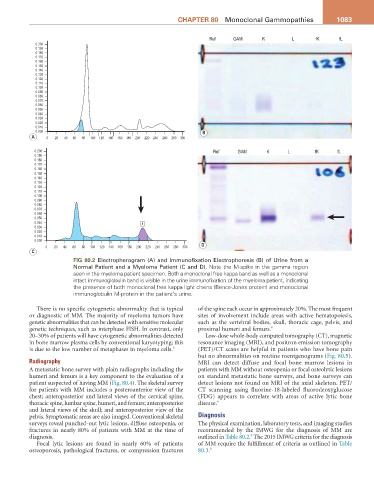
Fig 80.2 Electropherogram (A) and Immunofixation Electrophoresis (B) of Urine from a
Normal Patient and a Myeloma Patient (C and D). Note the M-spike in the gamma region
seen in the myeloma patient specimen. Both a monoclonal free kappa band as well as a monoclonal
intact immunoglobulin band is visible in the urine immunofixation of the myeloma patient, indicating
the presence of both monoclonal free kappa light chains (Bence-Jones protein) and monoclonal
immunoglobulin M-protein in the patient’s urine.
There is no specific cytogenetic abnormality that is typical of the spine each occur in approximately 20%. The most frequent
or diagnostic of MM. The majority of myeloma tumors have sites of involvement include areas with active hematopoiesis,
genetic abnormalities that can be detected with sensitive molecular such as the vertebral bodies, skull, thoracic cage, pelvis, and
genetic techniques, such as interphase FISH. In contrast, only proximal humeri and femurs. 6
20–30% of patients will have cytogenetic abnormalities detected Low-dose whole-body computed tomography (CT), magnetic
in bone marrow plasma cells by conventional karyotyping; this resonance imaging (MRI), and positron emission tomography
is due to the low number of metaphases in myeloma cells. 6 (PET)/CT scans are helpful in patients who have bone pain
but no abnormalities on routine roentgenograms (Fig. 80.5).
Radiography MRI can detect diffuse and focal bone marrow lesions in
A metastatic bone survey with plain radiographs including the patients with MM without osteopenia or focal osteolytic lesions
humeri and femurs is a key component to the evaluation of a on standard metastatic bone surveys, and bone surveys can
patient suspected of having MM (Fig. 80.4). The skeletal survey detect lesions not found on MRI of the axial skeleton. PET/
for patients with MM includes a posteroanterior view of the CT scanning using fluorine-18-labeled fluorodeoxyglucose
chest; anteroposterior and lateral views of the cervical spine, (FDG) appears to correlate with areas of active lytic bone
thoracic spine, lumbar spine, humeri, and femurs; anteroposterior disease. 6
and lateral views of the skull; and anteroposterior view of the
pelvis. Symptomatic areas are also imaged. Conventional skeletal Diagnosis
surveys reveal punched-out lytic lesions, diffuse osteopenia, or The physical examination, laboratory tests, and imaging studies
fractures in nearly 80% of patients with MM at the time of recommended by the IMWG for the diagnosis of MM are
8
diagnosis. outlined in Table 80.2. The 2015 IMWG criteria for the diagnosis
Focal lytic lesions are found in nearly 60% of patients; of MM require the fulfillment of criteria as outlined in Table
osteoporosis, pathological fractures, or compression fractures 80.3. 8