
Page 1122 - Clinical Immunology_ Principles and Practice ( PDFDrive )
P. 1122
1086 Part eight Immunology of Neoplasia
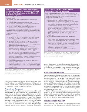
TABLE 80.2 exams, tests, and imaging TABLE 80.3 iMWg Criteria for the
Studies recommended by the international Diagnosis of Multiple Myeloma
Myeloma Working group for the Diagnosis Clonal bone marrow plasma cells ≥10% or biopsy-proven bony or
of Multiple Myeloma extramedullary plasmacytoma and any one or more of the following
CRAB (hyperCalcemia, Renal, Anemia, and Bone) features and
1. History and Physical Examination myeloma defining events (MDEs):
2. Routine Testing
• Complete blood count with differential and peripheral blood 1. Evidence of end-organ damage that can be attributed to the
underlying plasma cell proliferative disorder, specifically:
smear review.
• Chemistry panel including calcium and creatinine. • Hypercalcemia: serum calcium >0.25 mmol/L (>1 mg/dL) higher
than the upper limit of normal or >2.75 mmol/L (>11 mg/dL)
• Serum protein electrophoresis, immunofixation electrophoresis.
• Nephelometric quantitation of immunoglobulins. • Renal insufficiency: creatinine clearance <40 mL per minute or
serum creatinine >177 µmol/L (<2 g/dL [20 g/L])
• Routine urinalysis, 24-hour urine collection for proteinuria,
electrophoresis, and immunofixation electrophoresis. • Anemia: hemoglobin value of >20 g/L below the lowest limit of
• Quantification of both urine M-component level and albuminuria. normal, or a hemoglobin value <10 g/dL (100 g/L)
3. Bone Marrow Testing: Obtain an aspirate plus trephine biopsy with • Bone lesions: one or more osteolytic lesions on skeletal
radiography, CT, or PET/CT. If bone marrow has <10% clonal
testing for cytogenetics, fluorescent in situ hybridization (FISH), and
immunophenotyping. plasma cells, more than one bone lesion is required to
4. Imaging distinguish from solitary plasmacytoma with minimal marrow
involvement
• Bone survey including spine, pelvis, skull, humeri, and femurs. 2. Any one or more of the following biomarkers of malignancy
• The IMWG now recommends the use of low-dose whole-body
CT (LDWBCT) or MRI in the workup of smoldering multiple (MDEs):
• 60% or greater clonal plasma cells on bone marrow examination
myeloma (SMM) and solitary plasmacytoma.
• The IMWG now recommends that one of PET/CT, LDWBCT, or • Serum-involved uninvolved free light chain ratio of 100 or
greater, provided the absolute level of the involved light chain is
MRI of the whole body or spine be done in all patients with
suspected smoldering myeloma, with the exact imaging modality at least 100 mg/L (a patient’s “involved” free light chain—either
kappa or lambda—is the one that is above the normal reference
determined by availability and resources.
• Clear evidence of one or more sites of osteolytic bone range; the “uninvolved” free light chain is the one that is
typically in, or below, the normal range)
destruction (≥5 mm in size) seen on CT (including LDWBCT) or
PET/CT does fulfill the criteria for bone disease in multiple • More than one focal lesion on MRI that is at least 5 mm or
greater in size
myeloma and should be regarded as meeting the CRAB
(hyperCalcemia, Renal, Anemia, and Bone) requirement, whether Source: Rajkumar SV, Dimopoulos MA, Palumbo A, Blade J, Merlini G, Mateos MV,
or not the lesions can be visualized on skeletal radiography. et al. International Myeloma Working Group updated criteria for the diagnosis of
• Increased uptake on PET/CT alone is not adequate for the multiple myeloma. Lancet Oncol. 2014;15(12):e538–48. doi: 10.1016/S1470-
diagnosis of multiple myeloma; evidence of underlying osteolytic 2045(14)70442-5. PubMed PMID: 25439696.
bone destruction is needed on the CT portion of the
examination.
• Bone densitometry studies are not sufficient to determine the
presence of multiple myeloma.
• The IMWG no longer recommends the presence of osteoporosis
or vertebral compression fractures in the absence of lytic lesions abnormal plasma cell immunophenotype, and abnormalities on
as being sufficient evidence of bone disease for purposes of the imaging (MRI or PET/CT). 11,12 Patients with SMM should not
diagnostic criteria. be treated but instead closely monitored with follow-up every
3–4 months with treatment initiated upon progression to MM. 11
Source: Rajkumar SV, Dimopoulos MA, Palumbo A, Blade J, Merlini G, Mateos MV,
et al. International Myeloma Working Group updated criteria for the diagnosis of
multiple myeloma. Lancet Oncol 2014;15(12):e538–48. doi: 10.1016/S1470- NONSECRETORY MYELOMA
2045(14)70442-5. PubMed PMID: 25439696.
Approximately 3% of patients with MM have no M-protein in
the serum or urine based on immunofixation electrophoresis at
the time of diagnosis. Patients with myeloma who have normal
serum and urine immunofixation electrophoresis as well as a
the underlying plasma cell disorder, and no amyloidosis. SMM normal serum FLC ratio are considered to have true nonsecretory
is differentiated from MGUS by the quantity of M-protein in myeloma. Of these, approximately 85% will have M-protein that
serum (≥3 g/dL), urine (≥500 mg/24 h), and/or the percentage can be detected in the cytoplasm of the neoplastic plasma cells
of clonal bone marrow plasma cells (10–60%). by immunohistochemistry; however, they have impaired secretion
of this protein. The other 15% do not have immunoglobulin
Prognosis and Management detectable in the plasma cells. 6
Patients with SMM will progress to symptomatic MM or AL Patients with true nonsecretory myeloma need to be monitored
amyloidosis at an approximate rate of 10%/year for the first 5 mainly on the basis of imaging tests and bone marrow studies.
years, 3%/year for the next 5 years, and 1–2%/year for the fol- Patients with nonsecretory MM are not at risk for myeloma
lowing 10 years. The majority of patients with SMM will progress, kidney as long as light chains cannot be detected in the urine,
11
with a median time to progression of 4.8 years. The following but they are at risk for other complications of MM. 6
factors are associated with an increased risk of progression:
abnormal free light chain κ/λ ratio (<0.125 or >8.0), bone marrow OLIGOSECRETORY MYELOMA
clonal plasma cells 50–60%, serum M-protein ≥3 g/dL (or progres-
sive increase in M-protein level), IgA SMM, immunoparesis, Approximately 5–10% of patients with MM have oligosecretory
high-risk genetic features, increased circulating plasma cells, myeloma at the time of diagnosis as defined by the absence of