
Page 1127 - Clinical Immunology_ Principles and Practice ( PDFDrive )
P. 1127
ChaPter 80 Monoclonal Gammopathies 1091
thickening, easy bruising, and subcutaneous nodules or plaques. of AL amyloidosis. Kidney or liver biopsy is positive for amyloid
Amyloidosis may also be directly associated with a bleeding deposition in over 90% of cases; however, a high success rate
diathesis. Approximately 10% of patients have coexisting MM. 24 can also be achieved by less invasive procedures, such as abdominal
fat pad aspirate, rectal biopsy, bone marrow biopsy, or skin
KeY CONCePtS biopsy. 24
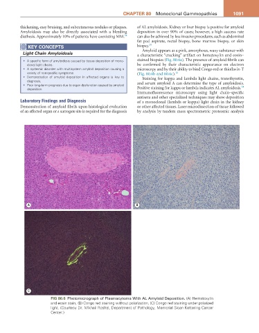
Amyloid appears as a pink, amorphous, waxy substance with
Light Chain Amyloidosis a characteristic “cracking” artifact on hematoxylin and eosin–
• A specific form of amyloidosis caused by tissue deposition of mono- stained biopsies (Fig. 80.6a). The presence of amyloid fibrils can
clonal light chains. be confirmed by their characteristic appearance on electron
• A systemic disorder with multisystem amyloid deposition causing a microscopy and by their ability to bind Congo red or thioflavin-T
variety of nonspecific symptoms. (Fig. 80.6b and 80.6c). 24
• Demonstration of amyloid deposition in affected organs is key to Staining for kappa and lambda light chains, transthyretin,
diagnosis.
• Poor long-term prognosis due to organ dysfunction caused by amyloid and serum amyloid A can determine the type of amyloidosis.
24
deposition. Positive staining for kappa or lambda indicates AL amyloidosis.
Immunofluorescence microscopy using light chain–specific
antisera and other specialized techniques may show deposition
Laboratory Findings and Diagnosis of a monoclonal (lambda or kappa) light chain in the kidney
Demonstration of amyloid fibrils upon histological evaluation or other affected tissues. Laser microdissection of tissue followed
of an affected organ or a surrogate site is required for the diagnosis by analysis by tandem mass spectrometric proteomic analysis
A B
C
Fig 80.6 Photomicrograph of Plasmacytoma With AL Amyloid Deposition. (A) Hematoxylin
and eosin stain, (B) Congo red staining without polarization, (C) Congo red staining under polarized
light. (Courtesy Dr. Mikhail Roshal, Department of Pathology, Memorial Sloan Kettering Cancer
Center.)