
Page 118 - Clinical Immunology_ Principles and Practice ( PDFDrive )
P. 118
102 Part one Principles of Immune Response
third form of the proteasome, the thymoproteasome, has alternative
forms of the β 5 subunit, β 5t . The thymoproteasome is selectively Endoplasmic reticulum Trafficking to Phagosomes or
expressed in the thymus and permits the use of alternative peptides TAP-independent Golgi and surface endosomes
for positive selection of CD8 T cells during development (Chapter peptides from
8). This leads to a broader CD8 TCR repertoire with diminished secreted proteins Endocytosed or
reactivity to self peptides expressed in the periphery. phagocytosed
Release from antigens
Import of Antigenic Peptides Into the Endoplasmic Reticulum class I loading
and Final Trimming complex
After antigenic or self peptides are generated by the proteasome MHC class I Cross-processing
in the cytosol, they are imported into the ER for potential binding heavy chain TAP
to newly synthesized MHC class I molecules. This transport β m Cytosol
2
activity, which must cross the ER membrane, is mediated by a
protein heterodimer termed TAP (for transporter associated with
antigen processing). The TAP heterodimer consists of TAP1 and Tapasin Proteasome
TAP2, each with 6 transmembrane domains, which within the Unfolded or
ER form a transmembrane channel. The adenosine triphosphate TAP-dependent partially degraded
(ATP) binding domains are on the cytosolic side of the endo- peptides protein
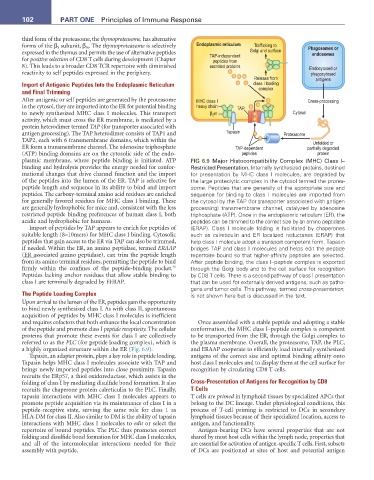
plasmic membrane, where peptide binding is initiated. ATP FIG 6.9 Major Histocompatibility Complex (MHC) Class I–
binding and hydrolysis provides the energy needed for confor- Restricted Presentation. Internally synthesized proteins, destined
mational changes that drive channel function and the import for presentation by MHC class I molecules, are degraded by
of the peptides into the lumen of the ER. TAP is selective for the large proteolytic complex in the cytosol termed the protea-
peptide length and sequence in its ability to bind and import some. Peptides that are generally of the appropriate size and
peptides. The carboxy-terminal amino acid residues are enriched sequence for binding to class I molecules are imported from
for generally favored residues for MHC class I binding. These the cytosol by the TAP (for transporter associated with antigen
are generally hydrophobic for mice and, consistent with the less processing) transmembrane channel, catalyzed by adenosine
restricted peptide binding preferences of human class I, both triphosphate (ATP). Once in the endoplasmic reticulum (ER), the
acidic and hydrophobic for humans. peptides can be trimmed to the correct size by an amino peptidase
Import of peptides by TAP appears to enrich for peptides of (ERAP). Class I molecule folding is facilitated by chaperones
suitable length (8–10mers) for MHC class I binding. Cytosolic such as calreticulin and ER localized reductases (ERAP) that
peptides that gain access to the ER via TAP can also be trimmed, help class I molecule adopt a transport competent form. Tapasin
if needed. Within the ER, an amino peptidase, termed ERAAP bridges TAP and class I molecules and helps edit the peptide
(ER associated amino peptidase), can trim the peptide length repertoire bound so that higher-affinity peptides are selected.
from its amino terminal residues, permitting the peptide to bind After peptide binding, the class I–peptide complex is exported
36
firmly within the confines of the peptide-binding pocket. through the Golgi body and to the cell surface for recognition
Peptides lacking anchor residues that allow stable binding to by CD8 T cells. There is a second pathway of class I presentation
class I are terminally degraded by EERAP. that can be used for externally derived antigens, such as patho-
gens and tumor cells. This pathway, termed cross-presentation,
The Peptide Loading Complex is not shown here but is discussed in the text.
Upon arrival to the lumen of the ER, peptides gain the opportunity
to bind newly synthesized class I. As with class II, spontaneous
acquisition of peptides by MHC class I molecules is inefficient
and requires cofactors that both enhance the local concentration Once assembled with a stable peptide and adopting a stable
of the peptide and promote class I peptide receptivity. The cellular conformation, the MHC class I–peptide complex is competent
proteins that promote these events for class I are collectively to be transported from the ER, through the Golgi complex to
referred to as the PLC (for peptide loading complex), which is the plasma membrane. Overall, the proteasome, TAP, the PLC,
a highly organized structure within the ER (Fig. 6.9). and ERAAP cooperate to efficiently load internally synthesized
Tapasin, an adapter protein, plays a key role in peptide loading. antigens of the correct size and optimal binding affinity onto
Tapasin helps MHC class I molecules associate with TAP and host class I molecules and to display them at the cell surface for
brings newly imported peptides into close proximity. Tapasin recognition by circulating CD8 T cells.
recruits the ERp57, a thiol oxidoreductase, which assists in the
folding of class I by mediating disulfide bond formation. It also Cross-Presentation of Antigens for Recognition by CD8
recruits the chaperone protein calreticulin to the PLC. Finally, T Cells
tapasin interactions with MHC class I molecules appears to T cells are primed in lymphoid tissues by specialized APCs that
promote peptide acquisition via its maintenance of class I in a belong to the DC lineage. Under physiological conditions, this
peptide-receptive state, serving the same role for class 1 as process of T-cell priming is restricted to DCs in secondary
HLA-DM for class II. Also similar to DM is the ability of tapasin lymphoid tissues because of their specialized location, access to
interactions with MHC class I molecules to edit or select the antigen, and functionality.
repertoire of bound peptides. The PLC thus promotes correct Antigen-bearing DCs have several properties that are not
folding and disulfide bond formation for MHC class I molecules, shared by most host cells within the lymph node, properties that
and all of the intermolecular interactions needed for their are essential for activation of antigen-specific T cells. First, subsets
assembly with peptide. of DCs are positioned at sites of host and potential antigen