
Page 116 - Clinical Immunology_ Principles and Practice ( PDFDrive )
P. 116
100 Part one Principles of Immune Response
Studies in both intact APCs and in vitro with purified recom-
binant class II and HLA-DM proteins suggest that DM selectively Targeting of Antigen into the MHC Class II
releases some peptides from class II molecules. Generally, these Processing Pathway
peptides possess low affinity for the class II molecule. Only those MHC class II molecules use several mechanisms to access self,
peptides that are stably bound to class II molecules will sustain tumor, or pathogen-derived proteins. In the case of extracellular
interactions with MHC class II and survive endosomal DM antigens, proteins can access endosomal compartments through
editing. These MHC class II complexes will be successfully receptor-mediated uptake, fluid phase uptake, or via internaliza-
exported to the cell surface, displayed at high density on the tion of cell surface molecules. The major pathways utilized will
APC and recruit CD4 T cells during an immune response. These vary with the type and the activation state of APCs. For DCs
DM-selected peptides are thus the targets of the focused CD4 and macrophages, calcium-dependent C-type lectins play a major
T-cell response. role in accessing exogenous protein antigens or pathogens. 27-29
The relationship between DM sensitivity and the affinity of These lectins recognize carbohydrate recognition domains (CRDs)
the peptide–class II complex is not absolute. However, it is specific for different sugar side structures on self or foreign
generally a good predictive feature of immunodominant peptides. proteins. Examples include O-linked or N-linked sugars on
In some cases, the size and diversity of the CD4 T-cell repertoire glycoproteins or O-linked sugars on glycolipid molecules. Among
can also determine the abundance of responding CD4 after the C-type lectins are those that recognize mannose-bearing
encounter with pathogens or vaccines, but the qualitative ability ligands (mannose receptor, SIGNR1, and langerin) and those
of a peptide to successfully or ineffectively recruit CD4 T cells that recognize complex sugars (dectin 1).
can be generally attributed to how stably the peptide binds to Receptors that have been implicated in antigen presentation
host HLA-class II molecules and thus DM editing. Importantly, include DEC205, expressed on lymphoid-resident DCs and DIR2
modification of this parameter by increasing the affinity of peptide (also known as Clec4) and its human orthologue DCIR2. Clec9A
binding to class II can enhance the immunogenicity of peptides, is a C-type lectin receptor that preferentially induces CD4
and thus can be used to enhance immunogenicity of vaccine T-follicular helper (Tfh) cells, which promote B-cell production
candidates. of isotype-switched high-affinity antibodies. Combined with
ligands for pattern recognition receptors or immunostimulatory
agents, such as CD40-specific antibodies, antigens delivered
HLA-DO through these cell surface proteins can activate CD4 T cells and
A final critical component of the MHC class II presentation induce protective immunity. Use of antibodies to these lectins,
pathway is HLA-DO (DO) in humans (H-2O in mice). The which allow precise targeting of specific subsets of APCs with
expression can be regulated independently of invariant chain antigen conjugates, has been exploited in vaccine design strategies
and HLA-DM. DO is unevenly distributed in MHC class II– to elicit the desired immune response to weakly immunogenic
positive cells. Factors influencing expression include cell lineage antigens, such as tumor antigens and HIV.
and state of activation. The differential expression of DO has In B cells, MHC-restricted antigen presentation is largely
important biological consequences because it inhibits the activity limited to those antigens taken up through the specific binding
of DM. The expression of DO in APCs can thus diminish CLIP of the somatically variable cell-surface Ig 30-32 (Fig. 6.8). Ig-mediated
release from class II, peptide loading of MHC class II and DM uptake of antigen is up to 10,000 times more efficient than fluid
editing of the final repertoire of class II presented peptides. phase uptake. Antigen capture by B cells within lymph node and
HLA-DM binds to DO in the same regions that it does the classic spleen follicles can involve direct access of soluble antigens
antigen-presenting class II molecule, but with a higher affinity (<70 kDa) delivered via afferent lymphatics to FDCs. For larger
and independently of peptide binding. HLA-DO can thus be membrane-associated antigens, viral aggregates, or immune
considered to be a high-affinity substrate mimic for HLA-DM.
DO initiates its interaction with DM within the ER, and
the two protein dimers are transported together into endosomal/
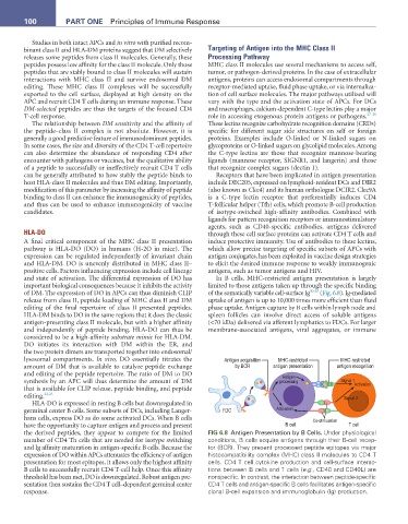
lysosomal compartments. In vivo, DO essentially titrates the Antigen acquisition MHC-restricted MHC-restricted
amount of DM that is available to catalyze peptide exchange by BCR antigen presentation antigen recognition
and editing of the peptide repertoire. The ratio of DM to DO Antigen
synthesis by an APC will thus determine the amount of DM processing Signal 1 Activation
that is available for CLIP release, peptide binding, and peptide
editing. 22,23 Signal 2
HLA-DO is expressed in resting B cells but downregulated in
germinal center B cells. Some subsets of DCs, including Langer- FDC Activation
hans cells, express DO as do some activated DCs. When B cells Co-stimulation
have the opportunity to capture antigen and process and present B cell T cell
the derived peptides, they appear to compete for the limited FIG 6.8 Antigen Presentation by B Cells. Under physiological
number of CD4 Th cells that are needed for isotype switching conditions, B cells acquire antigens through their B-cell recep-
and Ig affinity maturation in antigen-specific B cells. Because the tor (BCR). They present processed peptide epitopes via major
expression of DO within APCs attenuates the efficiency of antigen histocompatibility complex (MHC) class II molecules to CD4 T
presentation for most epitopes, it allows only the highest affinity cells. CD4 T cell cytokine production and cell-surface interac-
B cells to successfully recruit CD4 T-cell help. Once this affinity tions between B cells and T cells (e.g., CD40 and CD40L) are
threshold has been met, DO is downregulated. Robust antigen pre- nonspecific. In contrast, the interaction between peptide-specific
sentation then sustains the CD4 T cell–dependent germinal center CD4 T cells and antigen-specific B cells facilitates antigen-specific
response. clonal B-cell expansion and immunoglobulin (Ig) production.