
Page 1315 - Clinical Immunology_ Principles and Practice ( PDFDrive )
P. 1315
CHaPtEr 94 Assessment of Neutrophil Function 1277
–8
Buffer fMLF (10 M) granules, and lysosomal-associated membrane protein 3 (LAMP-3,
Distance (AU) Distance (AU) or CD63), stored in the azurophilic granules. During exudation,
the expression of CD11b and CD18 is increased over that observed
–300 –200 –100 0 100 200 300 –300 –200 –100 0 100 200 300 in peripheral neutrophils, whereas the expression of CD43
–100 (leukosialin) and CD62L is markedly reduced.
The antibody 7D5 recognizes an extracellular epitope of gp91 phox
0 phox
and can be used to identify surface expression of gp91 as well
100
Distance (AU) 200 granules. Flow cytometric analysis of neutrophils stained with
phox
as mobilization of latent pools of gp91
stored in the specific
7D5 can often be used to identify patients with X-linked chronic
300
400 granulomatous disease (CGD) (no 7D5 staining) and X-linked
chronic carriers of CGD (mosaic pattern of staining), particularly
500 in patients where the number of cells available for testing is limited.
The expression of surface antigens can also be used to assess
the responsiveness of neutrophils to particular ligands, such as
20
fMLF and LPS. As shown in Fig. 94.4, neutrophils isolated from
a patient who has a genetic defect in IL-1 receptor–associated
24
15 kinase-4 (IRAK-4) exhibit abnormal regulation of surface antigen
expression to LPS but exhibit normal regulation of surface antigen
expression to fMLF. Antigen expression can be upregulated
Velocity 10 because of translocation of latent antigen to the plasma membrane
or downregulated because of either internalization or shedding
of the antigen.
5
NEUTROPHIL DEGRANULATION
0 Clinical Indications and Implications
Random ν Directed ν Total ν Random ν Directed ν Total ν The proteases, acid hydrolases, and inflammatory mediators
released from storage granules in the neutrophils can mediate
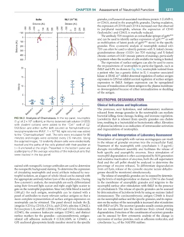
FIG 94.3 Analysis of Chemotaxis. In the top panel, neutrophils bacterial killing, tissue damage, healing, and immune regulation.
Lactoferrin that is released from specific granules can chelate
(1 µl of 2 × 10 cells/mL in Hanks balanced salt solution [HBSS] iron, resulting in a bactericidal or bacteriostatic effect. Elevation
6
with divalent cations) were added to the “Cell” well of EZ- of plasma lactoferrin is an indication of intravascular activation
TAXIScan and either buffer (left column) or formyl-methionyl- and degranulation of neutrophils.
–8
leucyl-phenylalanine (fMLF; 1 × 10 M, right column) was added
to the “Chemoattractant” well. The cells were incubated for 60 Principles and Interpretation of Laboratory Assessment
minutes and images were collected every 2.5 minutes. Using Stimulation of neutrophils with various secretagogues can result
the acquired images, 10 randomly chosen cells were electronically in the release of granular enzymes into the extracellular fluid.
tracked and the paths of the cells plotted with their position at Treatment of the neutrophils with cytochalasin b (5 µg/mL)
t = 0 anchored at the origin. Presented in the bottom panel are disrupts microfilament assembly and facilitates the release of
scattergrams of the average velocities of the individual cells that both specific and azurophilic enzymes. Since stimulation of
were tracked in the top panel. neutrophil degranulation is often accompanied by ROS generation
and oxidative inactivation of enzymes, both the cell supernatant
fluid and the cell pellet should be analyzed to determine the
stained with nonspecific isotype antibodies are used to determine percentage of enzyme released. To differentiate degranulation
the nonspecific background staining. To determine the expression from cell lysis, release of the cytosolic enzyme lactate dehydro-
of circulating neutrophils and avoid artifacts induced by neu- genase should be monitored simultaneously.
trophil isolation, an aliquot of whole blood can be stained with The release of azurophilic granules can be assessed by determin-
the appropriate antibody before lysis of the erythrocytes. During ing the levels of myeloperoxidase or elastase. CD63 is also found
flow cytometric analysis, the neutrophils are easily differentiated in the membrane of azurophilic granules and migrates to the
using their forward light scatter and right angle light scatter to neutrophil surface after stimulation with fMLF in the presence
gate on the neutrophil population. Since very little blood is needed of cytochalasin b. The release of specific granules can be assessed
(100 µL) for each antigen, neutrophils can be stained with a by determination of lactoferrin levels by using an enzyme-linked
panel of antibodies to many relevant surface antigens so that a immunoassay. The carcinoembryonic antigen CD66b is found
more complete representation of surface antigen expression on on the neutrophil surface and the specific granules, and its expres-
neutrophils can be obtained. The panel should include the β 2 sion on the surface of the neutrophils is increased after stimulation
integrins (CD11a, CD11b, CD11c, and CD18); selectins (CD62L); with fMLF or LPS. The secretory granules usually contain proteins
Fcγ receptors I, II, and III (CD64, CD32 and CD16); leukosialin that are translocated into the membrane from cytosol during
(CD43); the common leukocyte antigen (CD45); and distinct degranulation. Detection of the constituents of secretory granules
surface markers for the granules—carcinoembryonic antigen- can be assessed by flow cytometric analysis of the change in
related cell adhesion molecule 8 (CEACAM8, or CD66b), a expression of surface proteins, such as adhesion molecules, and
GPI-anchored glycoprotein family member stored in the specific cytochrome b 558 of the NADPH oxidase.