
Page 1319 - Clinical Immunology_ Principles and Practice ( PDFDrive )
P. 1319
CHaPtEr 94 Assessment of Neutrophil Function 1281
75
gp91 phox
gp67 phox
O 2 . (nmol/10 min/10 6 cells) gp22 phox Normal CGD CGD CGD Nonsense Missense Missense Missense
phox
gp47
50
– 25 p47 phox p67 phox p227 phox
gp91 phox CGD
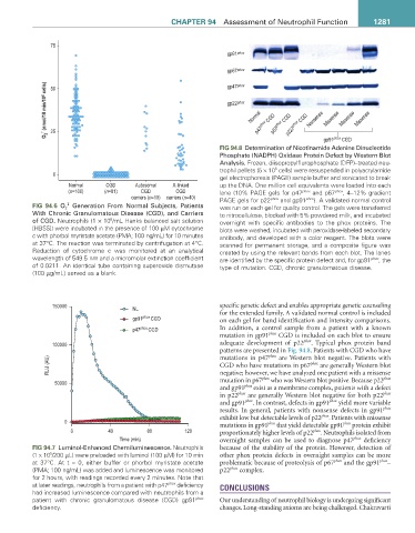
FIG 94.8 Determination of Nicotinamide Adenine Dinucleotide
Phosphate (NADPH) Oxidase Protein Defect by Western Blot
Analysis. Frozen, diisopropylflurophosphate (DFP)–treated neu-
trophil pellets (5 × 10 cells) were resuspended in polyacrylamide
6
0
gel electrophoresis (PAGE) sample buffer and sonicated to break
Normal CGD Autosomal X-linked up the DNA. One million cell equivalents were loaded into each
(n=100) (n=81) CGD CGD lane (10% PAGE gels for p47 phox and p67 phox , 4–12% gradient
carriers (n=10) carriers (n=40) PAGE gels for p22 phox and gp91 phox ). A validated normal control
•
FIG 94.6 O 2 Generation From Normal Subjects, Patients was run on each gel for quality control. The gels were transferred
With Chronic Granulomatous Disease (CGD), and Carriers to nitrocellulose, blocked with 5% powdered milk, and incubated
of CGD. Neutrophils (1 × 10 /mL Hanks balanced salt solution overnight with specific antibodies to the phox proteins. The
6
[HBSS]) were incubated in the presence of 100 µM cytochrome blots were washed, incubated with peroxidase-labeled secondary
c with phorbol myristate acetate (PMA; 100 ng/mL) for 10 minutes antibody, and developed with a color reagent. The blots were
at 37°C. The reaction was terminated by centrifugation at 4°C. scanned for permanent storage, and a composite figure was
Reduction of cytochrome c was monitored at an analytical created by using the relevant bands from each blot. The lanes
wavelength of 549.5 nm and a micromolar extinction coefficient are identified by the specific protein defect and, for gp91 phox , the
of 0.0211. An identical tube containing superoxide dismutase type of mutation. CGD, chronic granulomatous disease.
(100 µg/mL) served as a blank.
150000 NL specific genetic defect and enables appropriate genetic counseling
for the extended family. A validated normal control is included
gp91 phox CGD on each gel for band identification and intensity comparisons.
p47 phox CGD In addition, a control sample from a patient with a known
mutation in gp91 phox CGD is included on each blot to ensure
100000 adequate development of p22 phox . Typical phox protein band
patterns are presented in Fig. 94.8. Patients with CGD who have
phox
RLU (AU) mutations in p47 are Western blot negative. Patients with
phox
are generally Western blot
CGD who have mutations in p67
negative; however, we have analyzed one patient with a missense
50000 mutation in p67 phox who was Western blot positive. Because p22 phox
and gp91 phox exist as a membrane complex, patients with a defect
in p22 phox are generally Western blot negative for both p22 phox
and gp91 phox . In contrast, defects in gp91 phox yield more variable
results. In general, patients with nonsense defects in gp91 phox
exhibit low but detectable levels of p22 phox . Patients with missense
0 phox phox
mutations in gp91 that yield detectable gp91 protein exhibit
0 40 80 120 proportionately higher levels of p22 phox . Neutrophils isolated from
Time (min) overnight samples can be used to diagnose p47 phox deficiency
FIG 94.7 Luminol-Enhanced Chemiluminescence. Neutrophils because of the stability of the protein. However, detection of
5
(1 x 10 /200 µL) were preloaded with luminol (100 µM) for 10 min other phox protein defects in overnight samples can be more
at 37°C. At t = 0, either buffer or phorbol myristate acetate problematic because of proteolysis of p67 phox and the gp91 phox –
(PMA; 100 ng/mL) was added and luminescence was monitored p22 phox complex.
for 2 hours, with readings recorded every 2 minutes. Note that
at later readings, neutrophils from a patient with p47 phox deficiency CONCLUSIONS
had increased luminescence compared with neutrophils from a
patient with chronic granulomatous disease (CGD) gp91 phox Our understanding of neutrophil biology is undergoing significant
deficiency. changes. Long-standing axioms are being challenged. Chakravarti