
Page 206 - Clinical Immunology_ Principles and Practice ( PDFDrive )
P. 206
186 PART ONE Principles of Immune Response
P propagating activation signals. The importance of normal ITK
CD45
SH3 (Phosphatase) signaling was recently shown in humans, where homozygous
mutation led to immunodeficiency resulting in death from
P SH2 domain EBV-associated lymphoproliferation.
CSK Kinase SH2 domain
(Kinase) SH3
Kinase P KEY CONCEPTS
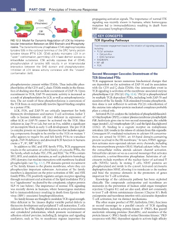
FIG 12.3 Model for Dynamic Regulation of LCK by Intramo- TCR Signaling Pathways
lecular Interaction Between an SH2 Domain and Phosphoty-
rosine. The transmembrane phosphatase CD45 dephosphorylates T-cell receptor engagement leads to the initiation of signaling cascades,
including:
tyrosine 505 in the carboxyl-terminus of the SRC family protein • PLCγ1 activation
tyrosine kinase (PTK) LCK. CD45 activity maintains LCK in an • Calcium flux
“open” conformation, permitting LCK kinase domain access to • DAG formation
intracellular substrates. CSK activity opposes that of CD45; • RAS/MAPK
phosphorylation of tyrosine 505 results in an intramolecular • NF- κB
interaction between the SH2 domain and phosphotyrosine. • PI3K
Inhibition of LCK kinase activity correlates with the “closed”
conformation (left). Second Messenger Cascades Downstream of the
TCR-Stimulated PTKs
TCR engagement incurs numerous biochemical changes that
phosphotyrosine present within ITAMs. Thus inducible phos- are dependent on the activation of ZAP-70 and its association
phorylation of the CD3 and ζ-chain ITAMs results in the forma- with the CD3 and ζ-chain ITAMs. One intermediate event in
tion of docking sites that mediate recruitment of ZAP-70. Upon TCR signaling is activation of the membrane-associated enzyme
7
recruitment to TCR, ZAP-70 enzymatic activity is increased as phospholipase Cγ1 (PLCγ1) (Fig. 12.4). PLCγ1 is phosphorylated
a result of phosphorylation by LCK as well as autophosphoryla- by multiple TCR-dependent PTKs, including both ZAP-70 and
tion. The net result of these phosphorylations is conversion of members of the Tec family. TCR-stimulated tyrosine phosphoryla-
the TCR from an enzymatically inactive ligand-binding complex tion alone is not sufficient to activate PLCγ1; relocalization of
to a potent PTK. the enzyme into adaptor-protein nucleated complexes probably
LCK, FYN, and ZAP-70 are critically important for both plays a critical role.
thymocyte development and mature T-cell activation. Jurkat T Activated PLCγ1 catalyzes the hydrolysis of phosphatidylinositol-
cells (a human leukemic cell line) deficient in expression of 4,5-bisphosphate (PIP2), a minor plasma membrane phospholipid.
either LCK or ZAP-70 cannot be activated via the TCR. Mice PIP 2 hydrolysis gives rise to two second messengers, the soluble
deficient in Zap-70 or Lck, but not in Fyn, exhibit a significant sugar inositol 1,4,5-trisphosphate (IP 3 ) and the lipid diacylglycerol
yet incomplete block in early T-cell development. The pre-TCR (DAG). IP 3 binding to a cognate receptor on endoplasmic
(a complex present on immature thymocytes that includes signal- reticulum (ER) results in the release of calcium from this organelle.
ing components thought to be similar to the TCR on mature T Consequent IP 3 -mediated reductions in calcium ER concentra-
cells) appears to require Src and Syk family PTKs to transduce tions are sensed by STIM1, an EF-hand domain–containing
8
signals. ZAP-70 deficiency and abnormal LCK function in humans protein localized in the ER membrane. In turn, STIM1 aggrega-
+
+
−
create a T , B , NK SCID. 5 tion activates store-operated calcium entry channels, including
In addition to SRC and SYK family PTKs, TCR engagement the transmembrane protein ORAI. Marked calcium influx from
results in the activation of a third family of cytosolic PTKs, the the extracellular milieu attends calcium channel activation.
6
Tec family, which includes TEC, ITK, and RLK. Tec PTKs contain Intracellular calcium serves as a second messenger that activates
SH2, SH3, and catalytic domains, as well as pleckstrin homology calcineurin, a serine/threonine phosphatase. Substrates of cal-
(PH) domains that mediate interactions with membrane-localized cineurin include members of the nuclear factor of activated T
phospholipids (see Fig. 12.2). PH domains permit recruitment cells (NFATs) family. In resting T cells, NFAT proteins are
of Tec family kinases to the plasma membrane, where they can phosphorylated and reside in the cytosol. Activated calcineurin
phosphorylate important substrates. Activation of Tec family dephosphorylates NFAT, allowing it to translocate to the nucleus
members is dependent on the prior activation of SRC and SYK and bind the response elements in the promoters of genes
family PTKs. ITK positively regulates antigen receptor signaling important for T-cell activation.
through recruitment and activation of the lipid modulator PLCγ Knowledge of the calcineurin pathway has been exploited
to a “signalosome” nucleated by the cytoplasmic adaptor protein clinically. The compounds cyclosporine and tacrolimus are
SLP-76 (see below). The importance of normal ITK signaling mainstays in the prevention of human solid-organ transplant
was recently shown in humans, where homozygous mutation rejection (Chapter 81) and are also used, albeit not commonly,
led to immunodeficiency resulting in death from Epstein-Barr to treat T cell–driven autoimmune diseases (Chapter 87). Both
virus (EBV)–associated lymphoproliferation. cyclosporine and tacrolimus interfere with calcineurin-dependent
Tec family kinases are thought to modulate TCR signal strength. T-cell activation, but via distinct mechanisms.
Mice deficient in Tec kinases display variable partial defects in The other major product of PIP 2 hydrolysis, DAG, functions
thymocyte development and peripheral T-cell maturation. T cells as second messenger to a parallel cascade of TCR signaling
deficient in Tec PTKs display alterations in actin polymerization intermediates, including protein kinase D (PKD), Ras guanyl
and cytoskeletal polarization, which promote recruitment of nucleotide releasing proteins (RasGRPs), and members of the
9
adhesion-related proteins, including β 2 integrins and signaling protein kinase C (PKC) family of serine/threonine kinases. PKD
mediators, such as Vav, to membrane regions important for cooperates with PKC-dependent signals to activate high-affinity