
Page 231 - Clinical Immunology_ Principles and Practice ( PDFDrive )
P. 231
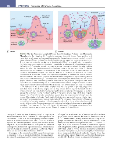
210          Part One  Principles of Immune Response


                                                                                                   Dendritic cell
                                               Intraepithelial                       Bacteria             Antimicrobial
                           Dendritic cell      lymphocyte                                                 peptides
             Lumen
                                                                              MAMPs      Dimeric IgA
                                               Goblet cell
                     M cell            T cell                        IgA+ cell
            Epithelium


                             B cell
             Lamina
             propria                     Cryptopatch
                   Peyer’s patch
                                                   Paneth cell
                                    LTi cell                         Mature isolated  Peyer’s patch
             Mesenteric                                              lymphoid follicle
             lymph node


         A  Prenatal                                         B  Postnatal
                       FIG 14.3  The Gut-Associated Lymphoid Tissue (GALT) Establishes Perinatal Host–Microbiota
                       Mutualism in the Intestine. (A) Prenatally, secondary lymphoid tissues (Peyer patches and
                       mesenteric lymph nodes) and cryptopatches develop by the spatiotemporal recruitment of lymphoid
                       tissue inducer (LTi) cells to sites of the developing intestine and supporting neurovascular structures.
                       This, in turn, stimulates the recruitment of dendritic cells (DCs), T cells, and B cells in preparation
                       for the immune response to the microbiota. Intraepithelial lymphocytes (IELs) seed the epithelium
                       before birth. (B) Postnatally, bacteria colonize the neonatal intestine immediately, initiating multiple
                       events that affect the development or functional maturation of the mucosa and GALTs. Shown
                       from left to right: Microbe-associated molecular patterns (MAMPs) sensed by pattern-recognition
                       receptors on intestinal epithelial cells and DCs adjacent to cryptopatches stimulate the further
                       recruitment of B cells and T cells, causing the cryptopatches to develop into mature isolated
                       lymphoid follicles. The isolated lymphoid follicles release immunoglobulin A (IgA)–producing plasma
                       cells, which are formed through T cell–dependent and independent interactions, into the lamina
                       propria. Microbes also cross the epithelium and enter the Peyer patch through M cells, from
                       which they are endocytosed by the DCs in the subepithelial dome. Antigen-loaded DCs in the
                       Peyer patch interact with local lymphocytes to induce T-cell differentiation and T cell–dependent
                       B-cell maturation in the germinal center (GC) to induce the development of IgA-producing plasma
                       cells that home to the lamina propria, where they release dimeric IgA for transport into the
                       intestinal lumen. DC-mediated luminal sampling of microbial products or transcytosis of bacteria
                       across the epithelium results in antigen loading of the lamina propria DCs, which then migrate
                       through the afferent lymphatics vessels (not shown) to a draining mesenteric lymph node to
                       induce differentiation of effector T cells that traffic to the lamina propria. Shown on the far right,
                       sensing of microbe-associated molecular patterns (MAMPs) stimulates the proliferation of intestinal
                       epithelial cells in crypts, resulting in their increased depth and, in the small intestine, increased
                       density of Paneth cells. This sensing also arms the intestinal epithelial cells for release of antimicrobial
                       peptides. (From Maynard CL, Elson CO, Hatton RD, Weaver CT. Reciprocal interactions of the
                       intestinal microbiota and immune system [Figure 1]. Nature 2012;489:231–241.)


                                                                                          +
                                                                       +
                                                                                    +
        (IFN-γ) and tumor necrosis factor-α (TNF-α) in response to   of NKp46  ILC3s and F4/80 CD11c  mononuclear cells in newborn
                                                                    3
        intracellular bacteria. ILC2s, similar to Th2 cells, express GATA3   pups.  In the normal intestine, ILC3s are the dominant source of
                                                                    12
        and secrete IL-13 and IL-5. ILC3s are arguably the most complex   IL-22.  The microbiota, acting on innate cells, including macro-
        subset of ILCs and can be subdivided into “LTi-like” ILC3s, which   phages and dendritic cells (DCs), induces expression of IL-23,
        populate the intestinal lymphoid follicles, and the more plastic   which, in turn, stimulates the production of IL-22 and/or IL-17
              +
        NKp46  ILC3s, which are distributed throughout the intestinal   by ILC3. IL-22 can induce the secretion of antimicrobial peptides,
        lamina propria. Both subsets express retinoic acid–related orphan   such as RegIIIγ, by intestinal epithelial cells to limit bacterial invasion,
                                                          +
        receptor (ROR)γt and produce IL-22 and IL-17, although NKp46    and within GALT to prevent systemic dissemination of resident
        ILC3s can also express Tbet and secrete IFN-γ. 12      microbes. Throughout life, ILC3 expressing Major Histocompat-
           The impact of the microbiota on ILC3 accumulation appears   ibility Complex (MHC) class II molecules (Chapter 5) on their
        to commence in utero, as transient colonization of pregnant germ-  cell surface help regulate the pool size and function of commensal-
        free mice with nonpathogenic Escherichia coli increases accumulation   specific CD4 T cells to prevent spontaneous inflammation. 13