
Page 287 - Clinical Immunology_ Principles and Practice ( PDFDrive )
P. 287
268 ParT TwO Host Defense Mechanisms and Inflammation
complexes from APCs via trogocytosis, capturing plasma mem- MDSCs are often associated with tumor progression and are
brane fragments from the immunological synapse and then believed to play an important role in the establishment of the
presenting them to other T cells in the absence of costimulatory immunosuppressive tumor environment. 26
signaling and possibly a signal-inducing apoptosis, such as Fas. DCs have also been demonstrated to have tolerogenic proper-
+
They may also suppress APCs in a manner similar to Foxp3 ties in certain circumstances. It is unclear if tolerogenic DCs are
Tregs by expression of CTLA-4. 22 of a stable lineage or, perhaps like Bregs, represent a particular
γδ T cells with a regulatory phenotype exist as a subset of state of differentiation. Antigen presentation by immature DCs
the epithelial γδ T cells, which can be found in mice. Mice may, indeed, be tolerogenic because of lack of costimulation, but
deficient in γδ T cells do not appropriately regulate responses antigen presentation by the same cells may be immunogenic once
to various pathogens. This inappropriate regulation manifests mature and expressing greater levels of costimulatory molecules.
as immunopathology in conjunction with the robust develop- Additionally, both pDCs found in the tumor microenvironment
+
ment of immunity. γδ T cell–deficient mice also show accelerated and CD103 conventional DCs in the lamina propria produce
autoimmune responses in models of systemic lupus erythema- the immune suppressive molecule indoleamine 2,3-dioxygenase
tosus (SLE) and spontaneously develop dermatitis when bred (IDO), which has been demonstrated to aid in the induction
on certain genetic backgrounds. Commonly, these conditions of pTreg cells. 27
are driven by αβ T cells, and γδ T cells will inhibit αβ T cells
predominantly in the local environment. In humans, who lack CLINICAL RELEVANCE OF REGULATORY T CELLS
an equivalent population of intraepithelial γδ cells, it is plau-
sible that this immune regulation is provided by other types of Abundant evidence strongly supports Tregs as key controllers
suppressive cells. 23 of self-tolerance, and Tregs of various subsets play an active role
NKT cells respond to CD1d, the nonclassic class I antigen- in the control of almost all types of physiological and pathological
presenting molecule, which binds glycolipids rather than peptides. immune responses, which also makes them suitable targets for
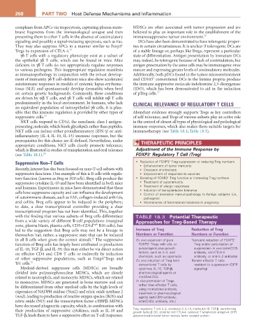
NKT cells can induce either proinflammatory (IFN-γ) or anti- immunotherapy (see Table 18.1; Table 18.3).
inflammatory (IL-4, IL-10, IL-13) immune responses, but the
prerequisites for this choice are ill defined. Nevertheless, under THEraPEUTIC PrINCIPLES
appropriate conditions, NKT cells clearly promote tolerance,
which is illustrated in studies of transplantation and oral tolerance Adjustment of the Immune Response by
+
(see Table 18.1). 24 FOXP3 Regulatory T Cell (Treg)
+
Suppressive Non–T Cells • Reduction of FOXP3 Treg suppression or reducing Treg numbers
• Enhancement of tumor immunity
Recently, interest has also been focused on non–T-cell subsets with • Clearance of infections
suppressive functions. One example of this is B cells with regula- • Improvement of responses to vaccines
+
tory function (known as Breg or B10 cells). Breg cells produce the • Boosting of FOXP3 Treg function or increasing Treg numbers
suppressive cytokine IL-10 and have been identified in both mice • Treatment of autoimmunity
and humans. Experiments in mice have demonstrated that these • Treatment of allergic responses
cells have suppressive capacity and can influence the development • Induction of transplantation tolerance
• Control of excessive immunopathology to foreign antigens (i.e.,
of autoimmune diseases, such as EAE, collagen-induced arthritis, pathogens)
and colitis. Breg cells appear to be induced in the periphery; • Maintenance of fetomaternal tolerance in pregnancy
to date, a clear transcriptional controller providing a clear
transcriptional program has not been identified. This, together
with the finding that various subsets of Breg cells differentiate TABLE 18.3 Potential Therapeutic
from a wide variety of different B-cell populations (marginal approaches for Treg-Based Therapy
zone, plasma blasts, plasma cells, CD5+CD1d high B10 cells), has
led to the suggestion that Breg cells may not be a lineage in Increase of Treg reduction of Treg
themselves but, rather, a suppressive state that can be induced Numbers or Function Numbers or Function
25
in all B cells when given the correct stimuli. The suppressive Ex vivo expansion of pure Transient reduction of FOXP3 +
+
function of Breg cells has largely been attributed to production FOXP3 Tregs with allo- or Treg and/or perturbation of
of IL-10, TGF-β, and IL-35; this may either be via direct action autoantigens plus growth suppression in vivo (anti-CD25
on effector CD4 and CD8 T cells or indirectly by induction factors, such as IL-2, and antibody, anti-CTLA-4
chemicals, such as rapamycin
antibody, or anti-IL-2 antibody)
+
of other suppressive populations, such as Foxp3 Tregs and Ex vivo induction of Treg from Render effector T cells
Tr1 cells. 25 conventional T cells by resistant to suppression (GITR
Myeloid-derived suppressor cells (MDSCs) are broadly cytokines (IL-10, TGF-β), signaling)
divided into polymorphonuclear MDSCs, which are closely pharmacological agents or
related to neutrophils, and monocytic MDSCs, which are related modified DCs
to monocytes. MDSCs are generated in bone marrow and can In vivo promotion of Tregs,
be differentiated from other myeloid cells by the high levels of rather than effector T cells,
using monoclonal antibody
expression of NADPH oxidase (Nox2) and nitric oxide synthase 2 treatment or pharmacological
(nos2), leading to production of reactive oxygen species (ROS) and agents (anti-CD3 antibody,
nitric oxide (NO) and the transcription factor c/EBPβ. MDSCs anti-CD40L antibody, etc.)
have decreased phagocytic capacity, which, in combination with
their production of suppressive cytokines, such as IL-10 and Treg, regulatory T cell; IL-2, interleukin-2; IL-10, interleukin-10; TGF-β, transforming
growth factor-β; DC, dendritic cell; CTLA-4, cytotoxic T lymphocyte antigen-4; GITR,
TGF-β, leads them to have a suppressive effect on T-cell responses. glucocorticoid-induced tumor necrosis factor receptor protein.