
Page 299 - Clinical Immunology_ Principles and Practice ( PDFDrive )
P. 299
280 ParT TwO Host Defense Mechanisms and Inflammation
Sensitization/immunization phase. Epidermal LCs and dermal during the sensitization phase, persist in the absence of antigens
DCs are responsible for carrying antigens from the affected skin and are readily capable of effector functions. They provide rapid
20
site to draining lymph nodes, where they present those antigens on-site immune protection to secondary antigen exposure.
to activate and expand antigen-specific T cells. To emigrate from Central memory T cells (T CM ) reside in lymph nodes and differ
the epidermis, activated LCs downregulate E-cadherin to detach from T RM , as they do not express high levels of skin-homing
from their surrounding keratinocytes. During migration to the receptors CLA and CCR4 or exhibit immediate robust responses
draining lymph node, LCs and DCs undergo a maturation process to antigen restimulation. T CM in lymph nodes or spleen can be
that stops antigen capture activity (by downregulation of antigen activated by secondary presentation of Ag by skin-draining LCs
capture receptors) and increases expression of T-cell interactive or DCs. Activated T CM enter the circulation, express skin-homing
molecules: MHC class I and II molecules, costimulatory molecules receptors, and extravasate into the affected skin site, where they
CD80 and CD86, and ICAM-1 (CD54). can contribute to the elicitation response. CD4 and CD8 T RM
Naïve T cells in the lymph nodes that express TCRs for and recruited T CM that interact with antigen-activated LCs and
presented antigens form long-term APC–T cell conjugates (on DCs in the dermis are further stimulated to proliferate in situ
the order of 30 minutes) during which T cells are programmed to amplify (i) effector cell numbers, (ii) cytokine and chemokine
to differentiate into functionally distinct T-cell subsets. These production, and (iii) further recruitment of inflammatory
new T cells leave the lymph node by downregulating their lymph leukocytes, which act in concert to eradicate microorganisms,
node–homing receptors, CCR7, and L-selectin (CD62L) and then chemically modified, or neoplastic cells.
upregulating the skin-specific homing receptor CLA and che-
mokine receptors CCR4 and CCR10 to enter the affected skin Skin Resident T-Cell Subpopulations
20
site. Unless antigen is present in skin for an extended period, In response to instructional cues from cutaneous DCs, antigen-
the sensitization phase does not cause an inflammatory reaction specific T cells differentiate into specialized subsets, each with
and therefore often goes unnoticed. functions that focus on eliminating specific types of pathogens
Elicitation/effector phase. In the dermis of resting skin, a large (Table 19.2). These presensitized, functionally committed T cells
number of resident memory T cells (T RM ), previously activated that home to skin remain in the dermis as long-lived T RM s. The
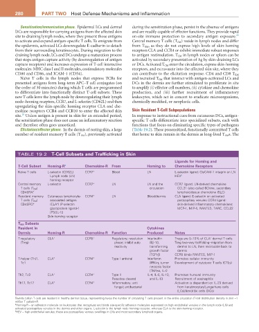
TABLE 19.2 T-Cell Subsets and Trafficking in Skin
Ligands for Homing and
T-Cell Subset Homing-r a Chemokine-r From Homing to Chemokine receptors
Naïve T cells L-selectin (CD62L) CCR7 + Blood LN L-selectin ligand: GlyCAM-1 integrin on LN
Lymph node (LN) HEV b
homing receptor
Central memory L-selectin CCR7 + LN LN and the CCR7 ligand: LN-derived chemokine
T cells (T CM ) circulation CCL21 (also called 6Ckine, secondary
CD45RA + lymphoid-tissue chemokine (SLC)
Resident memory Cutaneous lymphocyte- CCR4 + LN Blood/dermis CLA ligand: E-selectin on activated
T cells (T RM ) associated antigen postcapillary venules CCR4 ligand:
+
CD45RO + (CLA ) (P-selectin skin-derived inflammatory chemokines:
glycoprotein ligand-1 MCP-1, MIP-1, RANTES, TARC, CCL22
[PSGL-1])
Skin-homing receptor
T rM Subsets
resident in Cytokines
Dermis Homing-r Chemokine-r Function Produced Notes
+
T regulatory CLA + CCR5 + Regulatory; resolution Interleukin Tregs are 5–10% of CLA dermal T cells
(Treg) phase; inhibit auto (IL)-10, Treg two-way trafficking–migration from
reactivity transforming dermis to LN, then recirculate back to
growth factor dermis
(TGF)-β CCR5 binds RANTES, MIP-1
T-helper (Th)1, CLA + CCR4 + Type I antiviral Interferon Promotes cellular immunity
Tc1 (IFN)-γ, tumor Development of cytotoxic T cells (CTLs)
necrosis factor
(TNF)-α, IL-2
Th2, Tc2 CLA + CCR4 + Type II IL-4, IL-5, IL-10, Promotes humoral immunity
Parasites cleared and IL-13 Recruitment of eosinophils
Th17, Tc17 CLA + CCR4 + Inflammatory; anti Activation is dependent on IL-23 derived
fungal; antibacterial from keratinocytes/Langerhans cells
(LCs)/dendritic cells (DCs)
Twenty billion T cells are resident in healthy dermal tissue, representing twice the number of circulating T cells present in the entire circulation (T-cell distribution density in skin ≈1
million T cells/cm ).
2
a Homing-R – an adhesion molecule on leukocytes that recognizes and binds site-specific adhesion molecules expressed on high endothelial venules in the lymph node (LN) and
activated postcapillary venules in the dermis and other organs. L-selectin is the lymph node homing receptor, whereas CLA is the skin-homing receptor.
b HEV – high endothelial venules; these are postcapillary venous swellings in LNs and most secondary lymphoid organs.