
Page 37 - Clinical Immunology_ Principles and Practice ( PDFDrive )
P. 37
CHaPter 2 Organization of the Immune System 23
can enhance the growth and development of bone marrow
progenitor cells along multiple lineages in media containing IL-3, Mature Cells of the Immune System
IL-6, and GM-CSF. However, in the absence of other cytokines The mature cells of the immune system primarily arise from
or factors in serum, LIF has little effect on the growth and progenitor cells in bone marrow. They include both nonspecific
+
development of CD34 progenitors. TGF-β and IL-4 are potent and antigen-specific effector cells.
inhibitors of hematopoietic progenitor cell growth; yet they
enhance granulocyte development. Tumor necrosis factor-α Antigen-Presenting Cells
(TNF-α) inhibits the development of granulocytes, but it can The central player in both nonspecific and antigen-specific lines
potentiate IL-3 effects on hematopoietic progenitor cell of defense is the antigen-presenting cell (Chapter 6). In addition
proliferation. to their nonspecific effector functions, these cells are crucial for
Other cytokines have effects on the proliferation and dif- the development of specific immune responses. With maturation,
ferentiation of multipotent progenitors of hematopoietic and these cells enter the blood (Table 2.4) and circulate into the
lymphoid cells. GM-CSF and IL-3 promote development of tissues and organs.
granulocytes, macrophages, DCs, and erythrocytes. IL-6 partici- Antigen-presenting cells (APCs) are found in the solid
pates in the development of neutrophils, macrophages, platelets, lymphoid organs and skin (Chapter 19) at a frequency that varies
T cells, and B cells. Thrombopoietin signaling promotes stem from 0.1–1%. Specialized APCs in B-cell areas of lymph nodes
cell self-renewal to increase transplantation success. 7 and spleen are termed follicular dendritic cells (FDCs). They trap
antigen–antibody complexes important in the generation and
Cytokines That Inhibit Hematopoietic Stem Cell Growth maintenance of memory B cells. FDCs do not express major
Cytokines produced by mature cells can downregulate hemato- histocompatibility complex (MHC) class II molecules as do other
poietic stem cell growth. Macrophage inflammatory protein-1α APCs. Instead, they have receptors for immunoglobulin G (IgG)
(MIP-1α) is an inhibitor of hematopoietic progenitor cell (FcγRI [CD64]) and complement component C3b (CR1 [CD35]),
proliferation. Other factors regulate stem cell growth through a respectively.
variety of mechanisms, including the promotion of terminal
differentiation (e.g., interferon-γ [IFN-γ] and TGF-β) or through Monocytes–Macrophages
the induction of apoptosis (e.g., TNF-α). When pathologic Monocyte–macrophage lineage cells exist in blood (~10% of
conditions exist, these cytokines can have adverse effects on leukocytes) primarily as monocytes, which are large 10- to 18-µm
hematopoietic and lymphoid cell development. cells with peanut-shaped, pale purple nuclei as determined by
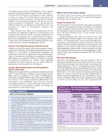
Wright staining (see Table 2.4). The cytoplasm, which is 30–40%
Cytokines Affecting Development and Differentiation of of the cell, is light blue and has azurophilic granules that resemble
Specific Cell Lineages ground glass with intracytoplasmic lysosomes. The cells express
Differentiation begins with the commitment of pluripotent stem MHC class II, CD14 (the receptor for lipopolysaccharide), and
cells to a specific lineage. Cytokines can have lineage-specific distinct Fc receptors (FcRs) for Ig. The latter include FcγRI (or
effects that act specifically at late stages of differentiation. CD64), which has a high affinity for IgG, and FcγRII (or CD32),
Erythropoietin regulates the later stages of erythrocyte differentia- which is of medium affinity and binds to aggregated IgG. FcγRIII
tion, whereas G-CSF induces granulocyte differentiation and (or CD16) has low affinity for IgG and is associated with
macrophage colony-stimulating factor (M-CSF) promotes
12
macrophage maturation. Cytokines that play an important role
in the growth and development of specific cell lineages are
described below under each cell type. TABLE 2.4 normal Distribution of White
Blood Cells in the Peripheral Blood of adults
KeY ConCePtS and Children
Cells of the Immune System aPProXIMate ranGe oF aBSoLUte
PerCentaGe CoUntS (no./µL)
• Pluripotent stem cells in bone marrow give rise to all lineages of the
immune system, platelets, and red blood cells. Children Children
• Development and regulation of cells of the immune system is associated Cell type adults (0–2 yr) adults (0–2 yr)
with programmed appearance of specific cell surface molecules called
“cluster of differentiation” (CD) markers and with responsiveness to Monocytes 4–13 400–1000 ND
selective cytokines. Dendritic cells 0.5–1 ND a 30–170 ND
• Mature cells of the immune system include antigen-presenting cells Granulocytes 35–73 2500–7500 1000–8500
(APCs); phagocytic cells, including neutrophils, eosinophils, and Lymphocytes 15–52 34–75 1450–3600 3400–9000
basophils; and lymphocytes, including T cells, B cells, and natural
killer (NK) cells, as well as other innate lymphoid cells. as % of Lymphocytes
• APCs include monocytes, macrophages, dendritic cells (DCs), B cells, T cells 75–85 53–84 900–2500 2500–6200
endothelial cells, epithelial cells, and adipocytes. They can direct the CD4 cells 27–53 32–64 550–1500 1300–4300
differentiation and function of both innate and acquired immune cells. CD8 cells 13–23 12–30 300–1000 500–2000
• Polymorphonuclear (PMN) granulocyte cells are important in the early B cells 5–15 06–41 100–600 300–3000
response to stress, tissue damage, and pathogens. They include Natural killer (NK) 5–15 03–18 200–700 170–1100
neutrophils, eosinophils, and basophils. cells
• Lymphocyte lineages have discrete subpopulations with specialized
functions. These include CD4 and CD8 T cells, B-1 and conventional a Not determined.
B-2 B cells, and NK and other innate lymphoid cells. CD4 T-helper Child data adapted from Shearer W, Rosenblatt H, Gelman R, et al. Lymphocyte
(Th) subsets include regulatory T cells (Tregs), Th17, and Tfh cells. subsets in healthy children from birth through 18 years of age: the pediatric AIDS
Clinical Trials Group P1009 study. J Allergy Clin Immunol 2003;12:973–80.