
Page 42 - Clinical Immunology_ Principles and Practice ( PDFDrive )
P. 42
28 Part one Principles of Immune Response
and TNFα and are the most heterogeneous. They also express Area of
CCR6 and CD117 and can be divided on the basis of expression stem cells
of the NCR Nkp44. The role of these cells in normal host function Erythropoiesis
island
and responses to chronic inflammatory stimuli and cancer is
under intensive study.
Bone
KeY ConCePtS
Tissues of the Immune System
• Stem cells proliferate and mature into effector cells in the primary
lymphoid organs, which include bone marrow and the thymus. Fat cells
• Mature immune cells reside in secondary lymphoid organs, where Lymphoid
additional maturation occurs and immune responses are generated. aggregate
• The spleen and lymph nodes comprise the systemic immune system,
which functions to protect the body from antigens in the lymphatic Megakaryocyte
drainage and the bloodstream.
• The mucosal immune system (respiratory, gastrointestinal, and genital)
and the skin and adipose tissue have distinguishing features that
differentiate the immune system at these sites from those of the A
systemic immune system.
• Mucosal sites include the mucosa-associated lymphoreticular tissue
(MALT).
• Commensal organisms at mucosal surfaces are an important
component of the immune response at these sites.
MAJOR LYMPHOID ORGANS
The primary lymphoid organs are sites where lymphocytes
differentiate from stem cells and then proliferate and mature
into effector cells. In humans, from birth to old age, these func-
tions are carried out only in bone marrow and the thymus.
Bone Marrow
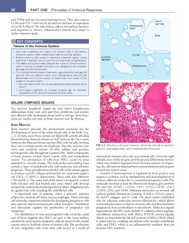
Bone marrow provides the environment necessary for the
development of most of the white blood cells of the body (Fig.
2.2). At birth, most bone cavities are filled with actively dividing
blood-forming elements known as “red” marrow. By 3 to 4 years, B
however, the tibia and femur become filled with fat cells, limiting
their role in hematopoietic development. The ribs, sternum, iliac FIG 2.2 Structure of bone marrow, showing islands of erythro-
crest, and vertebrae remain 30–50% cellular and produce poiesis, granulopoiesis, and scattered lymphocytes.
1
hematopoietic cells throughout a person’s life. Main components
of bone marrow include blood vessels, cells, and extracellular interactions between stem cells and stromal cells. Given the right
matrix. The production of cells from HSCs occurs in areas stimuli, most of the progeny proliferate and differentiate further,
separated by vascular sinuses. The walls of the surrounding sinus which may result in migration from the bone marrow. In migrat-
contain a layer of endothelial cells with endocytic and adhesive ing, the cells become detached from stromal elements and progress
properties. These specialized endothelial cells of the sinuses appear toward the central sinus.
to produce type IV collagen and laminin for structural support Control of hematopoiesis is regulated by both positive and
via CXCL-12 (SDF-1) interactions. These cells also elaborate negative cytokines, and by upregulation and downregulation of
CSFs and IL-6. The outer wall of the sinus is irregularly covered various adhesion molecules in committed progenitor cells. The
with reticular cells that branch into areas where cells develop molecules involved include the fibronectin receptor, glycoproteins
and provide anchors by producing reticular fibers. Megakaryocytes IIb and IIIa, ICAM-1 (CD54), LFA-1 (CD11, CD18), LFA-3
lie against this wall, touching the endothelial cells. (CD58), CD2, and CD44. Adhesion molecules on stromal cell
A functional unit of marrow, called a spheroid, contains surfaces include fibronectin, laminin, ICAM-1 (CD54), types I,
adipocytes, stromal cell types, and macrophages. These reticular III and IV collagen, and N-CAM. The most clearly established
cell networks compartmentalize the developing progenitor cells role for adhesion molecules involves fibronectin, which allows
into separate microenvironments called hematons. Osteoblasts erythroid precursors to bind to stromal cells and thus facilitates
and osteoclasts regulate the production of progenitor cell progression from erythroblast to reticulocyte. Molecular signals
expansion. 3 important for the HSC niche include N-cadherin, which regulates
The distribution of stem and progenitor cells across the radial osteoblastic interactions with HSCs; WNT/B catenin signals,
axis of bone suggests that HSCs are next to the bone surface, which are important for the self-renewal of HSCs; VEGF, which
whereas the more mature progenitor cells are nearer to the central is important for coupling osteoblasts with vascular endothelial
venous sinus to facilitate release of mature cells. The production cells; and PDE2, which is an inflammatory mediator that can
of new progenitor cells from stem cells occurs as a result of increase HSC numbers.