
Page 43 - Clinical Immunology_ Principles and Practice ( PDFDrive )
P. 43
CHaPter 2 Organization of the Immune System 29
CD 45 Lymphs Monos Seg neutro Macrophage Cortical
epithelium
Epithelium Capsule
Lymphoblasts Imm myel (+) selection
Hassall
Myeloblasts corpuscle
NRBC
Cortex Nurse cell (–) selection
Side scatter Dendritic cells Corticomedullary
FIG 2.3 Flow cytogram of Normal Human Bone Marrow junction
Based on CD45 Expression and Side Scatter. Most of the Medullary
major hematopoietic populations can be delineated. In this Pre-T epithelium
example 1.5% are red blood cell precursors (NRBCs), 1.5% are from BM Medulla
lymphoblasts, 3% are mature lymphocytes (Lymphs), 3% are CD4 + CD8 +
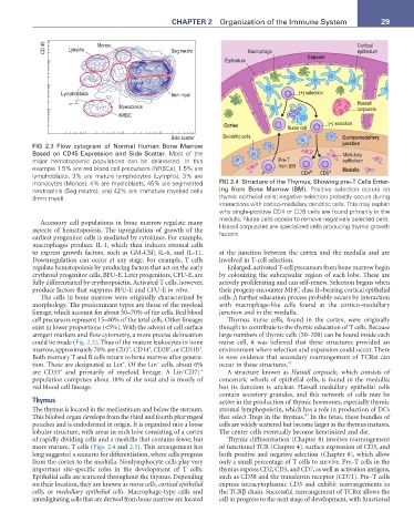
monocytes (Monos), 4% are myeloblasts, 45% are segmented FIG 2.4 Structure of the Thymus, Showing pre–T Cells Enter-
neutrophils (Seg neutro), and 42% are immature myeloid cells ing from Bone Marrow (BM). Positive selection occurs on
(Imm myel). thymic epithelial cells; negative selection probably occurs during
interactions with cortico-medullary dendritic cells. This may explain
why single-positive CD4 or CD8 cells are found primarily in the
medulla. Nurse cells appear to remove negatively selected cells.
Accessory cell populations in bone marrow regulate many Hassall corpuscles are specialized cells producing thymic growth
aspects of hematopoiesis. The upregulation of growth of the factors.
earliest progenitor cells is mediated by cytokines. For example,
macrophages produce IL-1, which then induces stromal cells
to express growth factors, such as GM-CSF, IL-6, and IL-11. at the junction between the cortex and the medulla and are
Downregulation can occur at any stage. For example, T cells involved in T-cell selection.
regulate hematopoiesis by producing factors that act on the early Enlarged, activated T-cell precursors from bone marrow begin
erythroid progenitor cells, BFU-E. Later progenitors, CFU-E, are by colonizing the subcapsular region of each lobe. These are
fully differentiated by erythropoietin. Activated T cells, however, actively proliferating and can self-renew. Selection begins when
produce factors that suppress BFU-E and CFU-E in vitro. their progeny encounter MHC class II–bearing cortical epithelial
The cells in bone marrow were originally characterized by cells. A further education process probably occurs by interaction
morphology. The predominant types are those of the myeloid with macrophage-like cells found at the cortico-medullary
lineage, which account for about 50–70% of the cells. Red blood junction and in the medulla.
cell precursors represent 15–40% of the total cells. Other lineages Thymus nurse cells, found in the cortex, were originally
exist in lower proportions (<5%). With the advent of cell surface thought to contribute to the thymic education of T cells. Because
antigen markers and flow cytometry, a more precise delineation large numbers of thymic cells (50–200) can be found inside each
could be made (Fig. 2.3). Thus of the mature leukocytes in bone nurse cell, it was believed that these structures provided an
+
+
+
+
marrow, approximately 70% are CD3 , CD14 , CD20 , or CD11b . environment where selection and expansion could occur. There
Both memory T and B cells return to bone marrow after genera- is now evidence that secondary rearrangement of TCRα can
−
+
tion. These are designated as Lin . Of the Lin cells, about 6% occur in these structures. 42
−
+
+
are CD33 and primarily of myeloid lineage. A Lin CD71 A structure known as Hassall corpuscle, which consists of
population comprises about 18% of the total and is mostly of concentric whorls of epithelial cells, is found in the medulla;
red blood cell lineage. but its function is unclear. Hassall medullary epithelial cells
contain secretory granules, and this network of cells may be
Thymus active in the production of thymic hormones, especially thymic
The thymus is located in the mediastinum and below the sternum. stromal lymphopoietin, which has a role in production of DCs
43
This bilobed organ develops from the third and fourth pharyngeal that select Tregs in the thymus. In the fetus, these bundles of
pouches and is endodermal in origin. It is organized into a loose cells are widely scattered but become larger as the thymus matures.
lobular structure, with areas in each lobe consisting of a cortex The center cells eventually become keratinized and die.
of rapidly dividing cells and a medulla that contains fewer, but Thymic differentiation (Chapter 8) involves rearrangement
more mature, T cells (Figs. 2.4 and 2.5). This arrangement has of functional TCR (Chapter 4), surface expression of CD3, and
long suggested a scenario for differentiation, where cells progress both positive and negative selection (Chapter 8), which allow
from the cortex to the medulla. Nonlymphocyte cells play very only a small percentage of T cells to survive. Pre–T cells in the
important site-specific roles in the development of T cells. thymus express CD2, CD5, and CD7, as well as activation antigens,
Epithelial cells are scattered throughout the thymus. Depending such as CD38 and the transferrin receptor (CD71). Pre–T cells
on their location, they are known as nurse cells, cortical epithelial express intracytoplasmic CD3 and exhibit rearrangements in
cells, or medullary epithelial cells. Macrophage-type cells and the TCRβ chain. Successful rearrangement of TCRα allows the
interdigitating cells that are derived from bone marrow are located cell to progress to the next stage of development, with functional