
Page 44 - Clinical Immunology_ Principles and Practice ( PDFDrive )
P. 44
30 Part one Principles of Immune Response
of cortical thymocytes to hormone-induced death probably
accounts for the involution, although human thymocytes are
Trabecula less sensitive to glucocorticosteroids compared with murine
thymocytes. However, an increase in steroids reduces immature
thymocyte numbers and enhances thymus involution. Recent
evidence suggests that active TCR rearrangements, and hence
T-cell development, continue in the adult thymus, albeit at a
Hassall corpuscle lower level than during childhood. There is an age-associated
decline in new T-cell production, such that by age 75 years, the
Medulla ability to make new T cells in humans is severely reduced.
Development of Hematopoietic and Lymphoid Cells
Although most of the key steps during the growth and develop-
ment of hematopoietic and lymphoid cells occur in bone marrow
Cortex and the thymus, additional maturation steps occur after the cells
leave those tissues. For example, monocytes and DC precursors
A migrate from blood vessels into tissues, where they mature into
macrophages and DCs, respectively. There is recent evidence for
a tissue-associated macrophage that is fetal in origin. Mast cells
and eosinophils also undergo further differentiation in resident
tissues. After leaving bone marrow and the thymus, B and T
cells undergo further maturation and memory cell development
in secondary lymphoid organs. There is strong evidence that
some T cells, particularly the γδ T cells residing in mucosal
epithelium, do not develop in the thymus.
SECONDARY LYMPHOID ORGANS
Secondary lymphoid organs are sites where mature lymphocytes
reside and where immune responses are generated. Secondary
lymphoid organs belong to either the systemic immune system
or the mucosal immune system. The systemic immune system,
which includes the spleen and lymph nodes, functions to protect
the body from antigens in the lymphatic drainage and circulating
in the bloodstream. The mucosal immune system responds to
B antigens that enter through mucosal epithelium and plays an
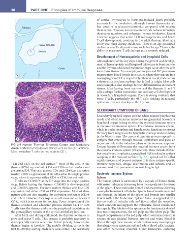
FIG 2.5 Human Thymus Showing Cortex and Medullary important role in the inductive phase of the immune response.
Areas. Cortical thymocytes are stained with an anti-CD1 antibody. Unique features differentiate the mucosal immune system from
Most medullary T cells do not express CD1. the systemic immune system (Chapter 20). These include efferent,
but not afferent, lymphatics, a specialized FAE involved in antigen
sampling at the mucosal surface (Fig. 2.6), specialized DCs that
rapidly process and present antigens to initiate antigen-specific
27
TCR and CD3 on the cell surface. Most of the cells in the immune responses, unique distribution and subsets, and an
thymus (85%) express both CD4 and CD8 on their surface and environment that promotes class switching to IgA.
are termed DP. They also express CD1 and CD69, an activation
marker. CD69 is expressed until the cell reaches the single-positive Systemic Immune System
stage, where it expresses either CD4 or CD8, but not both. Spleen
+
T cells are CD45RO at the DP stage into the single-positive The human spleen is surrounded by a capsule of fibrous tissue
stage. Before leaving the thymus, CD45RO is downregulated, with many trabeculae traversing from the capsule into the tissue
and CD45RA appears. The most mature thymus cells lose CD1 of the spleen. These trabeculae branch and anastomose, forming
expression and either CD4 or CD8 expression. Most of these a complex framework of lobules. Splenic blood vessels enter and
mature cells are also negative for activation molecules (CD38 exit through the hilum of the spleen and branch into smaller
and CD71). However, they acquire an adhesion molecule called vessels within the trabeculae. Splenic tissue is supported by a
CD44, which is necessary for homing. Upon completion of this fine network of reticular cells and fibers, called the reticulum,
thymus selection and education process, mature CD4 or CD8 which connects and supports the trabeculae, blood vessels, and
T cells leave the thymus and enter the peripheral circulation via the capsule. The lobules of the spleen can be functionally divided
the postcapillary venules at the cortico-medullary junction. into two compartments, the red pulp and the white pulp. The
After birth and during childhood, the thymus continues to largest compartment is the red pulp, which contains numerous
grow and select T cells. This process is probably necessary to venous sinuses situated between arteries and veins. Blood is
develop a fully normal repertoire. Before puberty, however, the filtered through these sinuses, which contain many macrophages
thymus begins to involute. The rapidly dividing cortex is the that phagocytose senescent red and white blood cells, bacteria,
first to atrophy, leaving medullary areas intact. The sensitivity and other particulate material. Other leukocytes, including