
Page 46 - Clinical Immunology_ Principles and Practice ( PDFDrive )
P. 46
32 Part one Principles of Immune Response
T cells play a key role in B-cell responses through CD40L and
other interactions. The signaling that occurs through this interac- Capsule
tion is central to B-cell activation and class switching. In addition
to activated B cells and CD4 T cells, the germinal center contains
FDCs and macrophages.
At the interface between the white pulp and the red pulp is
a region known as the marginal zone, which receives blood from
branches of central arterioles opening into this region. It contains
T cells, as well as subsets of macrophages and B cells. Marginal
zone (MZ) B cells are distinct from follicular B cells. They express
surface IgM and low levels of IgD and lack CD23. The initial Medulla
encounter of T cells and B cells with antigen occurs in the marginal Germinal
center
zone after blood enters through branches of the central arteriole.
Antigen presentation is enhanced by MZ B cells, which are Cortex
important in T cell–independent responses.
Lymph Nodes and Lymphatics
Lymph nodes occur as chains or groups located along lymphatic A
vessels. Lymph nodes exist in two major groups: those that drain
the skin and superficial tissues (e.g., cervical, axillary, or inguinal
lymph nodes) and those that drain the mucosal and deep tissues
of the body (e.g., mesenteric, mediastinal, and periaortic lymph
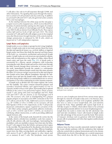
nodes). Lymph nodes are oval structures surrounded by adipose
tissue with an indentation at the region of a hilus, where blood
vessels enter and leave the node (Fig. 2.8). A lymph node is
surrounded by a fibrous capsule contiguous with trabeculae
traversing the node. Blood vessels and nerves, which enter through
the hilus, branch through these trabeculae to various parts of
the node. Immediately beneath the capsule is a subcapsular
(marginal) sinus. Afferent lymph vessels enter this sinus opposite
the hilus. DCs process antigen encountered in skin and migrate
into lymph nodes from afferent lymphatics through the sub-
capsular sinus and into the lymph node. Lymph nodes vary in
size, from being barely visible in an unstimulated state to several
centimeters in size when undergoing an active immune response.
A lymph node is divided into two major regions, the cortex
and the medulla. The cortex contains numerous primary and B
secondary lymphoid follicles, each approximately 0.5 mm in
diameter, similar to those in the spleen. Surrounding the lymphoid FIG 2.8 Human lymph node showing cortex, medullary areas,
follicles in the cortex is the paracortical region, which contains and germinal centers.
mostly T cells, along with some macrophages and DCs. Both
CD4 and CD8 T cells are present, as are macrophages and B
cells. The accessory cells, such as interdigitating DCs, present serves to carry lymphocytes derived from various tissue spaces
peptide antigens in association with MHC molecules to the TCR through the network of lymph nodes and to the thoracic duct.
on T cells to activate the T cells. Additional accessory molecules Lymphatic capillaries are lined with lymphatic epithelial cells
(e.g., B7 [CD80] or LFA-3 [CD58]) on the accessory cell and that serve as valves to move lymph fluid, cells, and nutrients
their ligands (CD28 or CD2, respectively) on the T cell provide around the body. These epithelial cells express high levels of
important costimulatory signals required for activation of the Toll-like receptor-4 (TLR4), which allows them to be activated
45
T cell. Other surface antigens, particularly adhesion molecules, after lipopolysaccharide delivery to increase lymphaniogenesis.
such as LFA-1 (CD18) and ICAM-1 (CD54), are involved in Lymph from the nodes is drawn into the left subclavian vein
stabilizing cellular interactions, as well as providing additional and back into circulation. Cancer cells found in lymph nodes
signals between cells. may take advantage of this system to seed the body. This system
In the center of the lymph node, beneath the cortex, lies the of transport develops early in gestation with both lymphatic
medulla, which is divided into medullary cords that contain T muscle cells for propulsion and valves that regulate unidirectional
cells, B cells, plasma cells, and macrophages. Surrounding the lymph flow.
medullary cords are medullary sinuses that drain into the hilus.
B and T cells migrate from the follicles and paracortical region Adipose Tissue
to the medulla. The Ig produced by the plasma cells drains into Adipose tissue has been recently extensively studied in light of
medullary sinuses that empty into the hilus. Efferent lymphatic the recent obesity epidemic with the realization that immune
vessels leave the hilus carrying lipids and antibodies, together cells play central roles in adipose homeostasis and in the chronic
with mature B and T cells that migrate to other tissues and inflammation in obesity. Macrophages are a central component.
act as memory B and T cells. The lymphatic vessel system They switch from the M2 to the M1 type in obesity. In lean