
Page 47 - Clinical Immunology_ Principles and Practice ( PDFDrive )
P. 47
CHaPter 2 Organization of the Immune System 33
adipose tissue, there are numerous Tregs and few CD8 T cells, Lamina propria Villous epithelium
which reverses during obesity when inflammation increases. 46
Mucosal Immune System
The mucosal immune system is located along the surfaces of
mucosal tissues (Chapter 20). Each mucosal immune system
consists of organized secondary lymphoid structures, termed
mucosa-associated lymphoreticular tissue (MALT), where inductive Germinal center Blood
vessels
immune responses occur, and more diffuse tissues, such as the
exocrine glands and lamina propria, where effector immune
47
responses occur. Some mucosal sites, such as the intestine and
lungs, have well-developed MALT. Others, such as the vaginal–
cervical mucosal surface, have minimally developed MALT.
MALT organization resembles lymph nodes with B cell follicles,
intervening T-cell zones and numerous APCs, such as DCs and
macrophages. Naïve T and B cells encounter antigen, become
activated, exit the tissue via efferent lymphatics, and migrate to
the local lymph nodes and then into the thoracic duct and the A
bloodstream. The cells home to effector sites, particularly the
lamina propria of various mucosal tissues. IELs, contained within
mucosal epithelium and the lamina propria located beneath the
epithelium, are responsible for effector functions. They occur
diffusely in mucosal tissues and lack the well-defined structure
of the organized mucosal immune system.
The homing of activated lymphocytes from one inductive
site to several mucosal surface effector sites has led to the concept
of a common mucosal immune system, although significant
48
compartmentalization remains in humans. Trafficking from
MALT to the lamina propria is well regulated. Expression of cell
surface molecules, such as sphingosine 1 phosphate (S1P),
MAdCAM-1, VLA-1, LFA-1, and VCAM-1; the cell surface
integrin, α4β7; and chemokines, such as CCR9, CCL25, CCR10,
and CCL28, are important in directing activated lymphocytes
49
to the lamina propria surface. The environment at the mucosal
surface is favorable for induction of IgA. Low-affinity IgA inhibits
commensal bacteria on mucosal surfaces. High-affinity IgA helps
neutralize microbial pathogens. B
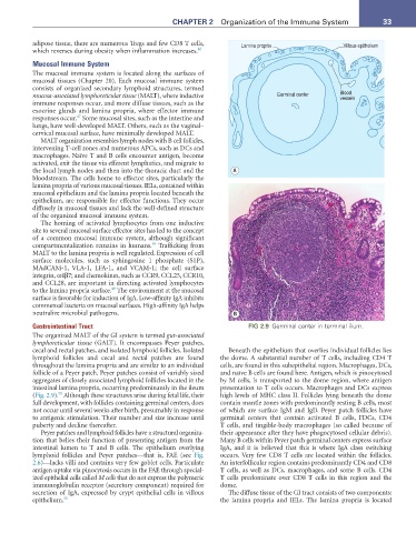
Gastrointestinal Tract FIG 2.9 Germinal center in terminal ilium.
The organized MALT of the GI system is termed gut-associated
lymphoreticular tissue (GALT). It encompasses Peyer patches,
cecal and rectal patches, and isolated lymphoid follicles. Isolated Beneath the epithelium that overlies individual follicles lies
lymphoid follicles and cecal and rectal patches are found the dome. A substantial number of T cells, including CD4 T
throughout the lamina propria and are similar to an individual cells, are found in this subepithelial region. Macrophages, DCs,
follicle of a Peyer patch. Peyer patches consist of variably sized and naïve B cells are found here. Antigen, which is pinocytosed
aggregates of closely associated lymphoid follicles located in the by M cells, is transported to the dome region, where antigen
intestinal lamina propria, occurring predominantly in the ileum presentation to T cells occurs. Macrophages and DCs express
50
(Fig. 2.9). Although these structures arise during fetal life, their high levels of MHC class II. Follicles lying beneath the dome
full development, with follicles containing germinal centers, does contain mantle zones with predominantly resting B cells, most
not occur until several weeks after birth, presumably in response of which are surface IgM and IgD. Peyer patch follicles have
to antigenic stimulation. Their number and size increase until germinal centers that contain activated B cells, FDCs, CD4
puberty and decline thereafter. T cells, and tingible-body macrophages (so called because of
Peyer patches and lymphoid follicles have a structural organiza- their appearance after they have phagocytosed cellular debris).
tion that belies their function of presenting antigen from the Many B cells within Peyer patch germinal centers express surface
intestinal lumen to T and B cells. The epithelium overlying IgA, and it is believed that this is where IgA class switching
lymphoid follicles and Peyer patches—that is, FAE (see Fig. occurs. Very few CD8 T cells are located within the follicles.
2.6)—lacks villi and contains very few goblet cells. Particulate An interfollicular region contains predominantly CD4 and CD8
antigen uptake via pinocytosis occurs in the FAE through special- T cells, as well as DCs, macrophages, and some B cells. CD4
ized epithelial cells called M cells that do not express the polymeric T cells predominate over CD8 T cells in this region and the
immunoglobulin receptor (secretory component) required for dome.
secretion of IgA, expressed by crypt epithelial cells in villous The diffuse tissue of the GI tract consists of two components:
epithelium. 51 the lamina propria and IELs. The lamina propria is located