
Page 478 - Clinical Immunology_ Principles and Practice ( PDFDrive )
P. 478
CHaPTEr 32 Approach to the Evaluation of the Patient With Suspected Immunodeficiency 459
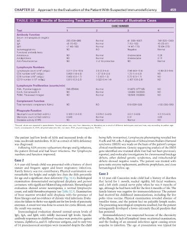
TABLE 32.3 results of Screening Tests and Special Evaluations of Illustrative Cases*
CaSE NuMBEr
Test 1 2 3 4
antibody Function
Serum immunoglobulin (mg/dL)
IgG 235 (208–686) Normal 41 (256–1067) 748 (520–1340)
IgA <8 (10–62) Normal 7 (12–103) 40 (25–81)
IgM <7 (43–183) Normal 14 (47–173) 78 (64–275)
Isohemagglutinins ND ND Negative Normal
Functional antibody tests
Antitetanus ND Normal Undetectable 0.28
Antidiphtheria ND Normal Undetectable 0.12
Anti-Pneumococcus ND 2 of 14 protective ND Normal
Lymphocyte Numbers
Lymphocyte count (×10 cell/µL) 0.217 (2.5–16.5) 2.8 (2.3–5.4) 2.56 (4.0–13.5) 10.60 (3.0–9.5)
3
CD4 number (×10 cell/µL) 0.009 (1.6–4.0) 1.27 (0.9–2.4) 1.72 (1.4–4.3) ND
3
3
CD8 number (×10 cell/µL) 0.008 (0.5–1.7) 1.5 (0.5–1.5) 0.73 (0.5–1.7) ND
3
CD19 number (×10 cell/µL) 0.010 (0.3–2.0) 0.023 (0.4–1.4) 0.107 (0.6–2.6) ND
Lymphocyte Proliferation (counts/min)
PHA, Phytohemagglutin 546 (55494) Normal 210973 (177195) ND
ConA, Concanavalin A ND Normal 124680 (143591) ND
PWM, Pokeweed mitogen ND Normal 162646 (138684) ND
Complement Function
Total hemolytic complement (U/mL) ND ND 618 (300–500) <100 (300–500)
Phagocyte Function
Neutrophil count (×10e3 cell/mL) 0.109 (1.0–9.0) 0.570 (1.5–8.5) 3.3 (1.0–8.5) 4.2 (1.5–8.5)
Monocyte count (×10e3 cell/mL) 0.546 Normal 0.11 0.65
Oxidative activity (DHR) ND Normal Normal ND
*Normal values are reported in parentheses. Normal values may vary from laboratory to laboratory as a result of different techniques used and may vary according to patient age.
ConA, concanavalin A; DHR, dihydrorhodamine; ND, not done; PHA, phytohemagglutinin; PVVM, .
The patient had low levels of ADA and increased levels of the being fully immunized. Lymphocyte phenotyping revealed low
toxic nucleotide metabolites. SCID as a result of ADA deficiency B cells and NK cells. A diagnosis of Schwachman-Bodian-Diamond
was diagnosed. syndrome (SBDS) was made on the basis of the patient’s unique
Following ADA enzyme replacement therapy and Ig infusions, clinical manifestations. Genetic sequencing analysis of the SBDS
the patient thrived and had fewer infections. Her lymphocyte gene identified one mutated allele that had not been previously
numbers and function improved. reported, and molecular investigations for chromosomal breakage
defects, other skeletal genetic syndromes, and mitochondrial
Case 2 defects showed negative results. The patient was treated with
A 4-year-old female child was presented with a history of short pancreatic enzyme replacement, antibiotic prophylaxis, and close
stature and frequent upper and lower respiratory infections. hematological follow-up.
Family history was not contributory. Physical examination was
remarkable for height and weight less than the fifth percentile Case 3
for age and a significant chest deformity (Fig. 32.6). Radiological A 12-year-old Caucasian male child had a history of diarrhea
examination demonstrated metaphyseal dysplasia and pectum that lasted for 1 month, nuchal rigidity, left facial weakness,
carinatum, with significant bilateral lung atelectasis. Hematological and a left sixth cranial nerve palsy when he was 8 months of
evaluation showed severe neutropenia, a normal lymphocyte age, although he had been well for the first 6 months of life. The
count, and mild thrombocytopenia (see Table 32.3). Examination family history was negative for immunodeficiency. The patient
of bone marrow revealed mild dysplasia with the presence of had received his childhood immunizations, including the live
three lineages and no evidence of chronic viral infections. Evalu- polio virus vaccine. Physical examination revealed absence of
ation for failure to thrive was significant for low levels of pancreatic tonsillar tissue, and the patient had no palpable lymph nodes.
enzymes. A sweat test was done to screen for cystic fibrosis, and The presenting neurological symptoms resolved, but the patient
the result was normal. subsequently developed a lower-extremity flaccid paralysis that
Immunological evaluation showed normal serum levels of has persisted.
IgG, IgA, and IgM, with mildly increased IgE levels. Specific Immunodeficiency was suspected because of the chronicity
antibody responses to childhood vaccines were protective against of the illness, the lack of lymphoid tissue on physical examination,
tetanus, diphtheria, and H. influenzae antigens, and to only two and the finding of an unusual infectious agent causing severe
of 14 pneumococcal serotypes were measured despite the child sequelae to infection. The age at presentation was typical for