
Page 587 - Clinical Immunology_ Principles and Practice ( PDFDrive )
P. 587
CHaPTEr 40 Autoantibody-Mediated Phenocopies of Primary Immunodeficiency Diseases 563
OVERVIEW OF PATHOPHYSIOLOGY dendritic cells (DCs), erythrocyte progenitors, and megakaryo-
cytes. The GM-CSF receptor is composed of two α and two β
The pathophysiology of infection susceptibility is generally subunits, which together bind two GM-CSF molecules with high
thought to result from a functional deficiency in the cytokine affinity and induce signal transduction and activator of transcrip-
that is being neutralized (Chapter 9). It is believed that a high- tion (STAT)–5 phosphorylation, nuclear translocation, and
titer autoantibody binds its respective cytokine target, thereby induction of master transcription factor PU.1. PAP was first
blocking downstream signaling and biological activity. For each described in 1958 by Rosen et al. as an idiopathic syndrome of
anticytokine–autoantibody pair, it has been demonstrated that respiratory failure, histopathologically characterized by alveolar
plasma or purified IgG from a patient with the anticytokine filling with acellular periodic acid-Schiff–positive proteinaceous
1
autoantibody prevents the activity of the targeted cytokine at the material. The pathogenesis of PAP has since been linked to
levels of signal transduction, gene transcription, and/or protein congenital or acquired defects in the GM-CSF signaling pathway.
expression. In the case of anti–IFN-γ autoantibodies, it appears The first clues to the etiological mechanism of PAP surfaced
−/−
8
that antibody levels may track with disease activity ; however, for in 1994 and 1995 when a GM-CSF and GM-CSF receptor
−/−
anti–GM-CSF autoantibodies, the results have been conflicting, β mice, respectively, demonstrated pulmonary disease that was
and in neither case has the question been rigorously studied. virtually identical to human PAP. Shortly thereafter, mechanisms
It is also possible, but not yet proven, that antibody-binding involving disruption of GM-CSF signaling were linked to PAP
1
avidity may influence the degree of disease severity as well. Thus in humans. Primary PAP results from mutations in either the
it may be possible to have high-titer, lower avidity anticytokine GM-CSF receptor subunits α or β and generally leads to severe
autoantibodies leading to a similar disease phenotype to low-titer, respiratory failure and usually presents early in life. Autoimmune
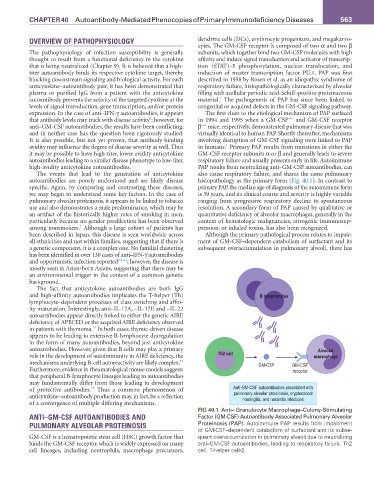
high-avidity anticytokine autoantibodies. PAP results from neutralizing anti–GM-CSF autoantibodies, can
The events that lead to the generation of anticytokine also cause respiratory failure, and shares the same pulmonary
autoantibodies are poorly understood and are likely disease histopathology as the primary form (Fig. 40.1). In contrast to
specific. Again, by comparing and contrasting these diseases, primary PAP, the median age of diagnosis of the autoimmune form
we may begin to understand some key factors. In the case of is 39 years, and its clinical course and severity is highly variable
pulmonary alveolar proteinosis, it appears to be linked to tobacco ranging from progressive respiratory decline to spontaneous
use and also demonstrates a male predominance, which may be resolution. A secondary form of PAP caused by qualitative or
an artifact of the historically higher rates of smoking in men, quantitative deficiency of alveolar macrophages, generally in the
particularly because no gender predilection has been observed context of hematologic malignancies, iatrogenic immunosup-
1
among nonsmokers. Although a large cohort of patients has pression, or inhaled toxins, has also been recognized.
been described in Japan, this disease is seen worldwide across Although the primary pathological process relates to impair-
all ethnicities and not within families, suggesting that if there is ment of GM-CSF–dependent catabolism of surfactant and its
a genetic component, it is a complex one. No familial clustering subsequent overaccumulation in pulmonary alveoli, there has
has been identified in over 130 cases of anti–IFN-γ autoantibodies
and opportunistic infection reported 2,9-12 ; however, the disease is
mostly seen in Asian-born Asians, suggesting that there may be
an environmental trigger in the context of a common genetic
background.
The fact that anticytokine autoantibodies are both IgG
and high-affinity autoantibodies implicates the T-helper (Th) B lymphocytes
lymphocyte–dependent processes of class switching and affin-
ity maturation. Interestingly, anti–IL-17A, –IL-17F, and –IL-22
autoantibodies appear directly linked to either the genetic AIRE
deficiency of APECED or the acquired AIRE deficiency observed
13
in patients with thymoma. In both cases, thymic-driven disease
appears to be leading to extensive B-lymphocyte dysregulation
in the form of many autoantibodies, beyond just anticytokine
autoantibodies. However, given that B cells may play a primary Alveolar
role in the development of autoimmunity in AIRE deficiency, the Th2 cell macrophage
13
mechanisms underlying B-cell autoreactivity are likely complex. GM-CSF GM-CSF
Furthermore, evidence in rheumatological mouse models suggests receptor
that peripheral B-lymphocyte lineages leading to autoantibodies
may fundamentally differ from those leading to development
14
of protective antibodies. Thus a common phenomenon of Anti-GM-CSF autoantibodies associated with
anticytokine–autoantibody production may, in fact, be a reflection pulmonary alveolar proteinosis, cryptococcal
meningitis, and nocardia infections
of a convergence of multiple differing mechanisms.
FIG 40.1 Anti– Granulocyte Macrophage–Colony-Stimulating
ANTI–GM-CSF AUTOANTIBODIES AND Factor (GM-CSF) Autoantibody Associated Pulmonary Alveolar
PULMONARY ALVEOLAR PROTEINOSIS Proteinosis (PAP). Autoimmune PAP results from impairment
of GM-CSF–dependent catabolism of surfactant and its subse-
GM-CSF is a hematopoietic stem cell (HSC) growth factor that quent overaccumulation in pulmonary alveoli due to neutralizing
binds the GM-CSF receptor, which is widely expressed on many anti–GM-CSF autoantibodies, leading to respiratory failure. Th2
cell lineages, including neutrophils, macrophage precursors, cell, T-helper cell-2.