
Page 610 - Clinical Immunology_ Principles and Practice ( PDFDrive )
P. 610
CHaPTEr 42 Urticaria, Angioedema, and Anaphylaxis 587
Eosinophil
C5b-9
ICAM1
t-PA
TNF-α IL-8
Bronchi
E-selectin
Histamine C3a
C5a
Mast cell
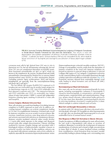
fiG 42.4 Immune Complex-Mediated Urticaria Initiated by Lodging of Antigenic Complexes
in Small Blood Vessels Followed by C3a and C5a Generation. This results in mast cell
degranulation and cytokine upregulation of adhesion molecules (intercellular adhesion molecule
[ICAM]-1, E-selectin) by tumor necrosis factor-α (TNF-α) and interleukin-8 (IL-8), which leads to
tissue recruitment of neutrophils and eosinophils and activation of tissue plasminogen activator
(t-PA).
cutaneous mast cells by IgG derived from CSU sera in vitro is (hypocomplementemic urticarial vasculitis syndrome [HUVS]).
dependent on C5a, but not all histamine-releasing IgG derived Damage to postcapillary venules results from the deposition of
from CSU sera are complement dependent on basophil assays. immune complexes in the vessel wall. Immune complexes are
Most functional autoantibodies are IgG1 or IgG3, which are formed on exposure to external (drug or infections) or internal
known to fix complement. By contrast, nonfunctional anti-FcεRI (collagen-like region of C1q) antigens. Complement activation
autoantibodies demonstrated by Western blot or enzyme-linked via the classical pathway leads to neutrophil chemotaxis through
immunosorbent assay (ELISA) in other autoimmune diseases cytokine expression and adhesion molecule activation (Fig. 42.4).
(including systemic lupus erythematosus [SLE] and bullous Proteolytic enzymes released from neutrophils damage vessel
pemphigoid) are generally from the noncomplement-fixing walls, leading to wheal formation and red blood cell (RBC)
subclasses IgG2 and IgG4. 10 extravasation.
The low-affinity IgE receptor FcεRII/CD23 expressed on B
lymphocytes and eosinophils may be another target antigen for Nonimmunological Mast-Cell Activation
an autoimmune response in CSU. Anti-CD23 antibodies have Skin mast cells can be activated nonimmunologically by many
11
been detected in patients with CSU. The interaction between agents, including neuropeptides (substance P, neuropeptide Y,
anti-CD23 antibodies and CD23 on eosinophils may induce vasoactive intestinal peptide, or somatostatin), calcitonin gene-
release of major basic protein 1 (MBP-1), a potent IgE-independent related protein, compound 48/80, and natural polyamines. Drugs
histamine-releasing agent, resulting in activation of mast cells (e.g., opiates, muscle relaxants, radiocontrast media, polymyxin
and basophils. B) can also cause dose-dependent nonimmunological urticaria
by activating membrane-associated G-coupled proteins. A classic
Immune Complex–Mediated Urticarial Rash clinical example is codeine-induced acute urticaria.
Mast-cell activation can result from binding of circulating immune
complexes to FcγRIII, expressed on mast cells. In addition, cir- Mast-Cell and Basophil Releasability in Urticaria
culating immune complexes can activate complement, leading In patients with CSU, dermal mast cells show a decreased activa-
to C3a and C5a anaphylatoxin formation. Urticarial rash caused tion threshold. Although CSU basophils are often hyporesponsive
by immune complexes can occur acutely in serum sickness–like to anti-IgE, they are paradoxically hyperresponsive to an as yet
reactions, transfusion reactions, some drug-induced urticarias, unidentified factor in normal human serum. 10
and urticaria associated with infectious or autoimmune diseases.
Immune complex–mediated urticarial rashes usually develop Skin Response to Mast-Cell Activation in Chronic Urticaria
1–3 weeks after initial exposure to the antigen and disappear Despite the existence of different pathways for mast-cell activation,
several weeks after antigen discontinuation. Chronic wheeling the end result is degranulation with release or secretion of
mediated by immune-complex damage and associated with a mediators. Histamine is crucial for the development of cutaneous
histological evidence of leukocytoclastic vasculitis is known as manifestations of urticaria and is found in high concentrations
urticarial vasculitis (see below). In this condition, urticaria can in tissue fluid of wheals. It causes localized redness resulting
be associated with systemic symptoms, such as fever, arthritis, or from vasodilatation, wheal formation from increased vascular
nephritis, especially the uncommon hypocomplementemic variant permeability leading to plasma leakage, and a surrounding axon