
Page 943 - Clinical Immunology_ Principles and Practice ( PDFDrive )
P. 943
910 Part Seven Organ-Specific Inflammatory Disease
SGPG GM1 GD1a GD1b GT1b GQ1b
Gal Gal Gal Gal NeuAc - Gal
Gal NeuAc - NeuAc - NeuAc -
cUA GlcNAc - GlcNAc GlcNAc GlcNAc
- GlcNAc GlcNAc NeuAc - -
- - - NeuAc - NeuAc -
- Gal NeuAc Gal NeuAc Gal NeuAc Gal NeuAc Gal NeuAc Gal
SO 3
Glc Glc Glc Glc Glc Glc
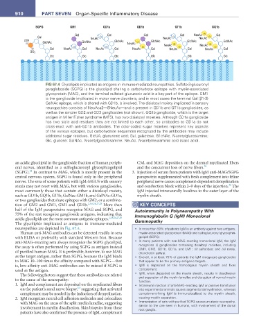
FIG 67.4 Glycolipids implicated as antigens in immune-mediated neuropathies. Sulfate-3-glucuronyl
paragloboside (SGPG) is the glycolipid sharing a carbohydrate epitope with myelin-associated
glycoprotein (MAG), and the terminal sulfated glucuronic acid is a key part of the epitope. GM1
is the ganglioside implicated in motor nerve disorders, and in most cases the terminal Gal (β1-3)
GalNAc epitope, which is shared with GD1b, is involved. The disialosyl moiety implicated in sensory
neuropathies consists of NeuAca2—8NeuAc—and is present in GD1b and GT1b gangliosides, as
well as the simpler GD2 and GD3 gangliosides (not shown). GQ1b ganglioside, which is the target
antigen in Miller Fisher syndrome (MFS), has two disialosyl moieties. Although GD1a ganglioside
has two sialic acid residues they are not linked to each other, so antibodies to GD1a do not
cross-react with anti-GD1b antibodies. The color-coded sugar moieties represent key aspects
of the various epitopes, but carbohydrate sequences recognized by the antibodies may include
additional sugar residues. GlcUA, glucuronic acid; Gal, galactose; G1cNAc, N-acetylglucosamine;
Glc, glucose; GalNAc, N-acetylgalactosamine; NeuAc, N-acetylneuraminic acid (sialic acid).
an acidic glycolipid in the ganglioside fraction of human periph- C3d, and MAG deposition on the dermal myelinated fibers
eral nerves, identified as a sulfoglucuronyl glycosphingolipid and the concurrent loss of nerve fibers. 29
28
(SGPG). In contrast to MAG, which is mostly present in the 3. Injection of serum from patients with IgM anti-MAG/SGPG
central nervous system, SGPG is found only in the peripheral paraprotein supplemented with fresh complement into feline
nerves. The sera of some patients with IgM-MGUS with sensory peripheral nerve causes complement-dependent demyelination
30
ataxia may not react with MAG, but with various gangliosides, and conduction block within 2–9 days of the injection. The
most commonly those that contain either a disialosyl moiety, IgM injected intraneurally localizes to the outer layer of the
such as GD1b, GQ1b, GT1b, GalNac-GM1b, and GalNAc-GD1a, myelin sheath.
or two gangliosides that share epitopes with GM2, or a combina-
tion of GM2 and GM1, GM1 and GD1b. 2,3,6,24,27,28 More than KeY COnCePtS
half of the IgM paraproteins recognize MAG and SGPG, and Autoimmunity in Polyneuropathy With
75% of the rest recognize ganglioside antigens, indicating that Immunoglobulin G (IgM) Monoclonal
acidic glycolipids are the most common antigenic epitopes. 2,3,6,24,27,28 Gammopathy
The glycolipids implicated as antigens in immune-mediated
neuropathies are depicted in Fig. 67.4. • In more than 50% of patients IgM is an antibody against two antigens,
Human anti-MAG antibodies can be detected readily in sera myelin-associated glycoprotein (MAG) and sulfoglucuronyl glycosphin-
with ELISA or preferably with standard Western blot. Because golipid (SGPG).
anti-MAG–reacting sera always recognize the SGPG glycolipid, • In many patients with non-MAG–reacting monoclonal IgM, the IgM
the assay is often performed by using SGPG as antigen instead recognizes (i) gangliosides containing disialosyl moieties, including
GM1, GM2, GD1b, GD1a, and LM1; (ii) sulfatides; and (iii) rarely,
of purified human MAG. It is preferable, however, to use MAG chondroitin sulfate.
as the target antigen, rather than SGPG, because the IgM binds • Overall, in at least 75% of patients the IgM recognizes gangliosides
to MAG 10–100 times the affinity compared with SGPG—that that appear to be the primary antigenic targets.
is, low-affinity anti-MAG antibodies can be missed if SGPG is • IgM is deposited on the homologous myelin sheath and fixes
used as the antigen. complement.
The following factors suggest that these antibodies are related • IgM, when deposited on the myelin sheath, results in disadhesion
and separation of the myelin lamellae and disruption of normal myelin
to the cause of the neuropathy: function.
1. IgM and complement are deposited on the myelinated fibers • Intraneural injection of anti-MAG–reacting IgM or passive transfusion
2,3
on the patient’s sural nerve biopsy, suggesting that activated into experimental animals causes segmental demyelination, whereas
complement may be needed in the induction of demyelination. complement-fixing IgM is immunolocalized to the myelin sheath,
2. IgM recognizes neural cell adhesion molecules and colocalizes causing myelin separation.
with MAG on the areas of the split myelin lamellae, suggesting • Immunization of cats with purified SGPG causes an ataxic neuropathy,
involvement in myelin disadhesion. Skin biopsies from these similar to the one seen in humans, with involvement of the dorsal
root ganglia.
patients have also confirmed the presence of IgM, complement