
Page 940 - Clinical Immunology_ Principles and Practice ( PDFDrive )
P. 940
CHaPter 67 Autoimmune Peripheral Neuropathies 907
CHRONIC INFLAMMATORY DEMYELINATING a relapsing course, with spontaneous remissions necessitating
POLYNEUROPATHY the need to periodically evaluate the usefulness of continuing
immunotherapeutic interventions. Because the demyelination
CIDP is the most common form of chronic APN with prevalence is multifocal, affecting spinal roots, plexuses, and proximal nerve
as high as 9/100 000. 2,3,11,12 It is also the most gratifying chronic trunks, 3,13 the clinicopathological picture may be variable,
autoimmune neuropathy because it is treatable in the majority accounting for the different manifestation of symptoms and
of the cases. It can be considered the chronic counterpart of GBS signs. 2,3,11,12 The most notable CIDP variants include the asym-
because of the various clinical, electrophysiological, histological, metrical, unifocal or multifocal, motor–sensory form (Lewis-
and laboratory similarities. CIDP differs from GBS predominantly Sumner syndrome); the pure motor form; the pure sensory form;
by its tempo, mode of evolution, prognosis, and responsiveness the sensory ataxic form; and the pure distal form.
to steroids. First described as a “steroid-responding relapsing
polyneuropathy,” CIDP shares with GBS a variety of common Diagnosis
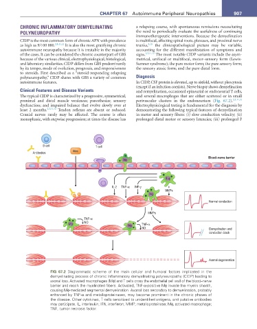
autoimmune features. In CIDP, CSF protein is elevated, up to sixfold, without pleocytosis
(except if an infection coexists). Nerve biopsy shows demyelination
Clinical Features and Disease Variants and remyelination, occasional epineurial or endoneurial T cells,
The typical CIDP is characterized by a progressive, symmetrical, and several macrophages that are either scattered or in small
proximal and distal muscle weakness; paresthesias; sensory perivascular clusters in the endoneurium (Fig. 67.2). 2,3,11,12
dysfunction; and impaired balance that evolve slowly over at Electrophysiological testing is fundamental for the diagnosis by
least 2 months. 2,3,11,12 Tendon reflexes are absent or reduced. demonstrating the following typical features of demyelination
Cranial nerves rarely may be affected. The course is often in motor and sensory fibers: (i) slow conduction velocity; (ii)
monophasic, with stepwise progression; at times the disease has prolonged distal motor or sensory latencies; (iii) prolonged F
B cell T cell
Mac
Antibodies
Blood–nerve barrier
Mφ
Mφ
IL-2 TNF-α INF-γ
MMPs
Mφ
Normal conduction
MMPs
TNF-α
Mφ NO TNF-α
Mφ
NO Demyelination and
conduction block
Axonal degeneration
FIG 67.2 Diagrammatic scheme of the main cellular and humoral factors implicated in the
demyelinating process of chronic inflammatory demyelinating polyneuropathy (CIDP) leading to
axonal loss. Activated macrophages (Mϕ) and T cells cross the endothelial cell wall of the blood–nerve
barrier and reach the myelinated fibers. Activated, TNF-α-positive Mϕ invade the myelin sheath,
causing Mϕ-mediated segmental demyelination. Axonal loss secondary to demyelination, probably
enhanced by TNF-α and metalloproteinases, may become prominent in the chronic phases of
the disease. Other cytokines, T cells sensitized to unidentified antigens, and putative antibodies
may participate. IL, interleukin; IFN, interferon; MMP, metalloproteinase; Mϕ, activated macrophage;
TNF, tumor necrosis factor.