
Page 941 - Clinical Immunology_ Principles and Practice ( PDFDrive )
P. 941
908 Part Seven Organ-Specific Inflammatory Disease
waves latencies; and (iv) conduction block with dispersion of transmigration across the blood–nerve barrier. Although T cells
the compound muscle action potentials. An associated axonal are not prominent overall, the few CD8 and CD4 cells found
loss is not unusual in the majority of CIDP cases. A variety of endoneurially have monoclonal or oligoclonal restrictions in
diagnostic criteria have been proposed to capture the most their T-cell receptor (TCR) repertoire, implying an antigen-driven
pertinent of the aforementioned features; the revised European T-cell response. 2,3,12,13
Federation of Neurological Societies/Peripheral Nerve Society Humoral factors seem to play a predominant role, especially
(EFNS/PNS) guidelines seem the most appropriate because they with the recent identification of target antigens at the nodes of
offer 81% sensitivity and 96% specificity to capture patients Ranvier in >10% of patients with CIDP, even though in the
more likely to respond to therapies. 2,3,11,12 Routine CSF testing majority of the patients, the pathogenic antigens remain elusive.
and nerve biopsy are not mandatory for the diagnosis, 2,3,11,12 but The beneficial effect of plasmapheresis provides indirect evidence
they can be helpful when the results of electrophysiological testing that some circulatory factors are pathogenic. The concept that
are not convincing, or there is a need to exclude hereditary and antibodies may be implicated dates back to >30 years when
vasculitic neuropathies. Other diseases causing neuropathy that complement-fixing IgG and IgM were found deposited on a
16
should be excluded include severe diabetes (although CIDP seems patient’s myelin sheath. Antibodies to glycolipids LM1, GM1,
more frequent in patients with diabetes), neoplasms, amyloidosis, or GD1b also have been seen in some patients, but less frequently
IgM paraproteinemia (IgG or IgA monoclonal gammopathy of than in GBS and more frequently than in controls. 2,3,11,12 Over-
undetermined significance (MGUS) can be seen in CIDP), whelming evidence accumulated in the last 5 years indicates that
myelomas, vasculitis, alcoholism, exposures to neurotoxic drugs, molecules associated with saltatory conduction at the nodes of
or family history of neuropathy. Ranvier may be more meaningful targets 3,4,10,17-19 because func-
tional blockade in these regions can best account for the rapid
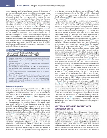
KeY COnCePtS improvement noticeable within days after plasmapheresis or
Autoimmunity in Chronic Inflammatory intravenous immunoglobulin (IVIG). Likely antigenic targets
include neurofascin-186, moesin, and gliomedin (at the node);
Demyelinating Polyneuropathy (CIDP) neurofascin-155 (NF155), contacting/Caspr 1 (CNTN1), and
• Activated macrophages are the predominant endoneurial cell, displacing connexins (at the paranode); and transient–axonal–glycoprotein-1
the Schwann cell cytoplasm, disrupting myelin, and lysing superficial or potassium channels (at the juxtaparanode). 3,4,10,17-19 Evidence
myelin lamellae. from proteomics, transfected cell lines, teased-nerve fibers,
• Complement-fixing immunoglobulin G (IgG) and IgM antibodies are immunocytochemistry, and enzyme-linked immunosorbent assay
deposited on the myelin sheath. (ELISA) has shown that IgG4 antibodies against paranodal NF155
• IgG antibodies to acidic glycolipids LM1, GM1, or GD1b and against and CNTN1 antigens are the most relevant and seemingly
the 28-kDa P0 myelin proteins are detected in the sera of some
patients. pathogenic antibodies that are currently detected in at least 10%
• There is upregulation of DR and B-7 costimulatory molecules in Schwann of patients. 17-19 Of interest, anti-NF155– and CNTN1-positive
cells and macrophages. patients appear to have a distinct clinical phenotype with more
• Serum IgG can induce conduction block when injected into rat nerves. severe disease, axonal involvement, tremors, sensory ataxia, and
• Up to 25% of patients with CIDP harbor specific antibodies against suboptimal response to IVIG 17-19 ; these patients, however, respond
antigens in the nodes of Ranvier; in 10% of them these antibodies better to anti–B cell therapies, such as rituximab.
have been identified as directed against neurofascin-155 and CASPR
causing conduction block. Overall, the immunopathogenetic scheme proposed for GBS
(see Fig. 67.1) is also appropriately applied to CIDP. Molecular
mimicry can be implicated in rare cases of CIDP associated with
Immunopathogenesis melanoma because the carbohydrate myelin epitopes GM2, GM3,
Because of immunopathological similarities to GBS and the and GD3 are also expressed on melanoma cells, and antibodies
20
relapsing–remitting EAN, in CIDP, activated T cells, macrophages, against melanoma cells react with myelin glycoproteins. The
complement, and autoantibodies seem to work in concert with axonal involvement that accompanies demyelination in both
each other to induce an immune attack against peripheral nerve CIDP and GBS, either acutely or after long-standing demyelin-
antigens 2,3,11,12 (as depicted in Fig. 67.1), but no triggering factors ation, is depicted in Fig. 67.2. The relevant nodal target antigens
have been identified so far. are presented in Fig. 67.3 separately 2,3,10 to highlight their rec-
The predominant endoneurial mononuclear cells in CIDP ognized importance in the field of antibody-mediated peripheral
are the macrophages that constitute the final effector cells associ- demyelination.
ated with demyelination because they express activation markers
probably induced by cytokines released by autoreactive T cells MULTIFOCAL MOTOR NEUROPATHY WITH
in situ or the circulation. These cells sequentially penetrate the CONDUCTION BLOCK
basement membrane of the Schwann cell, displace the cytoplasm,
and split the myelin lamellae, resulting in focal lysis of the myelin MMN is a distinct disease that, although rare with prevalence
sheath. 2,3,11,12 Macrophages as well as Schwann cells may serve of 0.6/100 000, should be recognized early because it is treatable.
as antigen-presenting cells (APCs) because they express human It affects males more than females and is more common in those
leukocyte antigen (HLA)-DR and costimulatory molecules B7-1 <50 years of age. MMN presents with progressive weakness,
(CD80) and B7-2 (CD86), whereas their counterreceptors atrophy, and areflexia that often begins in the hands and is
cytotoxic T lymphocyte antigen-4 (CTLA-4) and CD28 are prominent in distal muscle groups supplied by many individual
expressed on rare endoneurial CD4 T cells. 2,3,14 B7-2–deficient peripheral nerves (multifocal). 2,3,21 It differs from vasculitic
15
mice also develop CIDP. Elevated soluble adhesion molecules, neuropathy because it is slow and is painless because it affects
chemokines, cytokines, and metalloproteinases are detected in only the motor nerve fibers. It also differs from the motor variant
serum and CSF in CIDP, probably facilitating lymphoid-cell of CIDP because it is multifocal and asymmetrical. Because some