
Page 942 - Clinical Immunology_ Principles and Practice ( PDFDrive )
P. 942
CHaPter 67 Autoimmune Peripheral Neuropathies 909
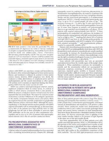
Target antigens at the Node of Ranvier: Explain rapid recovery neuropathy occurs in a setting of myeloma, plasmacytoma, or
Waldenström macroglobulinemia, the majority of patients with
paraproteinemic neuropathies do not have a lymphoproliferative
Axon disease, and the monoclonal gammopathy is of undetermined
JP PN N PN JP significance (MGUS). A benign monoclonal gammopathy may
22
occur in up to 1% of normal people >50 years of age. The
PN: Paranode N: Node JP: Juxtaparanode incidence increases to 1.7% above age 70 years and reaches up
Paranode Juxtaparanode to 6% above age 90 years. Monoclonal gammopathies, however,
ECM Myelin are 10 times more frequent in patients with polyneuropathy
gliomedin than in an age-matched control population, and almost 10% of
23
NF186 Tag1 patients with acquired polyneuropathy have MGUS. If these
NF155 Nav gammopathies are categorized into subclasses, the incidence of
Nav
Kv Kv polyneuropathy among patients with IgM monoclonal proteins
23,24
contactin Caspr2 can be as high as 50%, implying that almost 50% of patients
/Caspr ankyrinG with IgM MGUS may have or will develop polyneuropathy. At
βIVspectrin
Connexin 32, 29, 31.3 present, polyneuropathies with MGUS comprise 10% of patients
Axon with acquired neuropathy, 23-25 and paraproteinemic polyneu-
ropathy is a potentially treatable APN. 25
FIG 67.3 Main proteins in the nodal (N), paranodal (PN), and Patients with demyelinating polyneuropathy associated with
juxtaparanodal (JP) regions in the nodes of Ranvier implicated IgG or IgA MGUS are indistinguishable from those with CIDP; the
as antigens in acquired demyelinating neuropathies (Guillain-Barré paraprotein in such patients is coincidental and causally unrelated
syndrome [GBS] and predominantly chronic inflammatory to neuropathy. In contrast, the demyelinating polyneuropathy
demyelinating polyneuropathy [CIDP]). The two main antigenic associated with IgM MGUS is a distinct clinicopathologic entity,
targets in CIDP are neurofascin-155 and CASPR2 located in the and the IgM is considered pathogenic because it is often directed
paranodal regions; antibodies against these proteins are detected against myelin glycoproteins or glycolipids. 2,3,6,24,25
in the serum of 10% of patients with CIDP, resulting in conduction Some patients with paraprotein may have an associated
block and paranodal axonal changes that probably account for amyloidosis derived from the variable region of the Ig light chain,
resistance to IVIG. 4,13 primarily λ. When amyloidosis is present, the neuropathy is
painful, and the sensorimotor deficits are accompanied by
autonomic symptoms consisting of orthostatic hypotension,
patients with MMN can experience cramps and fasciculations, impotence, impaired gastric motility, or frequent diarrhea.
the disease is often erroneously diagnosed as lower motor neuron Amyloid neuropathy is difficult to treat; apart from symptomatic
disease, and treatment is delayed. In contrast to lower motor therapy (mostly opiates), immunosuppressive therapies, especially
neuron disease, however, MMN progresses very slowly, the with the various anti–B-cell agents, and bone marrow transplanta-
weakness is within distributions of peripheral nerves and not tion are largely applied.
multisegmental, and the cranial musculature is often spared.
CSF protein level is normal, an important finding that distin-
guishes MMN from the motor variant of CIDP. ANTIBODIES TO MYELIN-ASSOCIATED
MMN has a distinct electrophysiological criterion, namely, GLYCOPROTEIN IN PATIENTS WITH IgM M
multifocal conduction block in motor nerves. In contrast to
CIDP, in which sensory conduction block is also present, in MONOCLONAL GAMMOPATHIES OF
MMN the sensory conduction remains normal across the nerve UNDETERMINED SIGNIFICANCE
segments that have motor block. The reason for a selective motor POLYNEUROPATHY (ANTI-MAG NEUROPATHY)
involvement is unclear. Differences in the antigenic specificities
in the myelin components between motor and sensory fibers Most of these patients present with a sensory, large-fiber, demy-
are suspected because the ceramide composition of gangliosides elinating polyneuropathy that manifests as sensory ataxia. 2,3,24,26
differs between sensory and motor fibers. Although the immu- Other patients have a sensorimotor polyneuropathy with mixed
nopathology of the disease remains unclear, patients with MMN features of demyelination and axonal loss. CSF protein is often
respond remarkably well to immunotherapy, as discussed later. elevated. Nerve conduction studies have demonstrated slow
Up to 50% of the patients have high antibody titers to GM1 conduction velocity and a rather characteristic prolonged distal
ganglioside, but the role of these antibodies is uncertain. IgM motor and sensory latency indicative of distal demyelination.
GM1 antibodies can be also seen in other autoimmune neu- Sural nerve biopsy demonstrates a diminished number of myelin-
ropathies and in even up to 25% of patients with amyotrophic ated axons. On electron microscopy, there is splitting of the
lateral sclerosis (ALS) who do not have conduction block. outer myelin lamellae, linked to the presence of IgM deposits
in the same area of the split myelin sheath.
Sera from approximately 50% of these patients react with
POLYNEUROPATHIES ASSOCIATED WITH myelin-associated glycoprotein (MAG), a 100-kilodalton (kDa)
MONOCLONAL GAMMOPATHIES OF glycoprotein of the central and peripheral nerve myelin, as well
UNDETERMINED SIGNIFICANCE as other glycoproteins or glycolipids that share antigenic deter-
minants with MAG. 2,3,24,27,28 The antigenic determinant for the
A distinct subset of acquired polyneuropathies has been associated anti-MAG IgM resides in the carbohydrate component of the
3
with a circulating monoclonal protein (Chapter 80). Although MAG molecule. The anti-MAG IgM paraproteins coreact with