
Page 939 - Clinical Immunology_ Principles and Practice ( PDFDrive )
P. 939
906 Part Seven Organ-Specific Inflammatory Disease
Cytokines Barrier (Blood–brain)
(INF-γ, IL-1, IL-2, IL-6 (Blood–nerve)
TNF) APC
Integrins ICAM-1 IL-2 MHC B cell
MAC-1 MHC
TCR MHC-II
LFA-1
T cell MHC-I, II T cell Anti-MAG
INF-γ Autoantigen Anti-GQ1b
Anti-glycolipids
IL-2 IL-1(fever)
TNF-1
CR 1
ICAM-1 Fc
Viruses Mφ
MHC
MHC Mφ
CR 3
Mφ Mφ
TNF O 2
Schwann cell
IL-1 OH
Igs
MHC
CR 3 AG
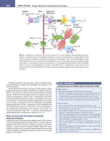
FIG 67.1 Sequence of events in the mechanisms of immune-mediated demyelinating polyneu-
ropathy. Cytokines lead to increased expression of major histocompatibility complex (MHC) class
I and intercellular adhesion molecules, allowing the sensitized T cells and macrophages to exit
the endothelial cell wall and traffic to the peripheral nerve. There they recognize myelin antigen
and induce a macrophage-mediated demyelination. The antigen-presenting cells (APCs; probably
Schwann cells or macrophages), in concert with MHC class II expression, interact with CD4 T
cells and lead to clonal expansion of B cells, producing antibodies against various peripheral nerve
antigens.
Molecular mimicry may also play a role in the Zika virus– KeY COnCePtS
associated GBS as antiglycolipid antibodies were found in 31%
of these patients. 8 Autoimmunity in Guillain-Barré Syndrome (GBS)
Molecular mimicry between epitopes of viral proteins (which Cellular Factors
trigger the disease) and myelin components may result in sensitiza- • The peripheral myelin or the Schwann cells are targets.
tion of cross-reactive T cells that may stimulate B cells to produce • Activated macrophages are the dominant endoneurial cells and lift
specific antibodies directed against myelin components or may the outermost myelin lamellae, lysing the superficial myelin sheaths.
recruit macrophages as effector cells. A combination of cellular • Peripheral blood lymphocytes exert myelinotoxic activity in vitro.
and humoral factors therefore seems to participate in the cause • Levels of interleukin-2 (IL-2) and soluble IL-2 receptors are increased
1-8
of the disease. Circulating cytokines triggered by the initiating during the acute phase of the disease and decline during recovery.
event (viruses or bacteria) could also upregulate intercellular Humoral Factors
adhesion molecule (ICAM)-1 expression on the endothelial cells • Serum exerts a complement-dependent demyelination in vitro.
and facilitate the entrance of activated T cells or antibodies to • Intraneural injections of serum from patients with acute GBS cause
the endoneurial parenchyma. It is relevant that ICAM-1 is demyelination and conduction block.
1-5
increased in patients with GBS. A scheme summarizing the • Immunoglobulin G (IgG), IgM, and membranolytic attack complex are
immunopathogenic mechanism is shown in Fig. 67.1. detected immunocytochemically on the patients’ nerves.
• High titers of IgG antibodies against peripheral nerve acidic glycolipids
Nodal and Paranodal Alterations and Specific (GM1, GQ1b) are detected in the sera of patients with acute motor
Antinodal Antibodies axonal neuropathy (AMAN) and Miller Fisher syndrome (MFS). The
GQ1b ganglioside is a specific antigen for MFS.
Antibodies to nodal and paranodal antigens seen in CIDP subsets, • There is a high incidence of antibodies to Campylobacter jejuni, and
as discussed later, have been also detected in a small number of GM1, with molecular mimicry between Campylobacter and nerve
patients with GBS in Europe. Antibodies against neurofascin, gangliosides.
ankyrin G, and contactin could potentially explain the acute • Injection of lipooligosaccharides extracted from C. jejuni cause AMAN
conduction block or the paranodal axonal degeneration and the and elicit GM1 antibodies in rabbits.
rapid reversibility or slow and incomplete recovery seen in several • Antiganglioside antibodies extracted from patients with GBS block
muscle action potentials in vitro.
patients with AMAN 1-5,9,10