
Page 1193 - Hall et al (2015) Principles of Critical Care-McGraw-Hill
P. 1193
830 PART 6: Neurologic Disorders
descriptive clinical syndrome rather than a disease-specific entity. The interestingly, normal subjects display higher brain metabolism during
most common causes include cardiac arrest, head trauma, severe brain wakefulness than in sleep in similar regions. Furthermore, thalamo-
10
infections, and various causes of thalamic necrosis. Vegetative states cortical disconnections can be identified using auditory and sensory
can also be seen in the terminal phase of degenerative illnesses such as external stimulations. 11
Alzheimer disease. Ambiguous terms for PVS such as apallic syndrome Some carefully performed studies identified patients with PVS who
and neocortical death should be avoided. 3,4 activate primary and associative cortex, depending on the complexity
Minimal conscious state can be diagnosed in patients displaying inconsis- and familiarity of the test stimuli. 12,13 Recent data obtained in patients
tent behavioral evidence of awareness of the environment, but they cannot with PVS reveal functional magnetic resonance imaging (fMRI) acti-
communicate and are unable to follow instructions reliably (Table 88-2). vation of the supplementary motor area (SMA) and parahippocampal
5
It describes a large group of patients who are different from vegetative areas in motor planning and visuospatial task stimulation paradigms,
patients in that they demonstrate some signs of awareness of their selves respectively. It is, however, unclear whether the identified activations
14
and their surroundings albeit inconsistently. The inconsistency may be very represent consciousness since no conclusions to the connectivity of
subtle or more pronounced wherein unless observed for a long periods of thalamocortical brain regions and larger neuronal networks can be
time, it is almost impossible for a clinician to determine otherwise. drawn. Similarly, brain plasticity with recovery of functional thalamo-
Akinetic mutism is a manifestation of hypothalamic or basal forebrain cortical connections and reestablishment of neuronal networks may
injury, which manifests as apparent depressed levels of consciousness in allow certain patients to regain consciousness after severe brain injury
a patient with well-formed sleep-wake cycles, with no external evidence and PVS. In recent years, we have gained much insight into which brain
9
of awareness or spontaneous motor activity. It is imperative in such areas seem necessary for conscious experience; however, future research
instances to have a rigorous neurological examination as well as a careful should broaden our knowledge about what form of brain activity in
review of neuroimaging and EEG. Abulia is a state in which the patient these areas confers consciousness.
is awake, has normal sleep wake cycle, and is very slow to respond to
stimuli. Mental function is usually normal when tested with sufficient
stimulation. It is secondary to bilateral frontal lobe disease and in severe IMPAIRED CONSCIOUSNESS:
instances may mimic or progress to akinetic mutism. AN ANATOMIC APPROACH
Clinical practice teaches that consciousness should be viewed as a con- Because coma is a sleep-like state, it is not surprising to find that the
tinuum between different pathological conditions and not as an all-or- neuroanatomy of coma is closely related to brain stem centers that regu-
none phenomenon, and that it is frequently difficult to identify definite late daily cycles of wakefulness and sleep: the reticular activating system
and consistent signs of conscious perception of environment and self in (RAS). In animals the RAS lies within the center of the brain stem,
patients with severe brain injuries. The latter limits the diagnostic certainty extending from the midbrain into the hypothalamus and thalamus. 15,16
1
of remaining brain function on clinical grounds since the identification Lesions in the pathways of the brain stem reticular formation or RAS
of consciousness relies purely on the deduction whether consciousness is have the greatest impact on changes in consciousness.
present or absent in a particular patient. To this end the assessment is fur- The RAS is a loosely organized core of polysynaptic neurons reaching
2
ther complicated by the fact that responses sought along different domains in the brain stem from the lower medulla through the paramedian pons
of awareness are summarized by the patient’s motor manifestations. to central midbrain. From here, the RAS projects into the diencepha-
Extensive and severe neuronal damage is causally related to loss of lon to several functionally related nuclei in the thalami (especially the
consciousness in patients with PVS, a finding supported by a study medial thalamus). Further cerebral projections are prominent to the
identifying decreased cortical radiolabeled flumazenil uptake (a benzo- inferomedial frontal lobes, but reach almost all parts of the cerebral
diazepine antagonist and neuronal marker). In corroboration, several cortex. The essential role of the RAS is arousal and maintenance of
6
studies consistently identified decreased cerebral glucose metabolism wakefulness; injury leads to reduction or failure of arousal. As the brain
and blood flow. However, brain regions with the most consistent stem RAS receives direct spinothalamic information, incoming sensory
7,8
decrease in cerebral glucose consumption in patients in PVS are the stimulations are not only projected to the sensory cortices, but are also
polymodal association areas of the frontal, temporal, and parietal lobes ; needed to activate the brain stem RAS for the maintenance of conscious-
9
ness. Within the brain stem and thalamus, the RAS is confined to rather
small anatomic areas; therefore, even small lesions can severely impair
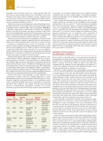
TABLE 88-2 Characteristics of the Persistent Vegetative State (PVS) arousal and consciousness. In contrast, RAS fibers are sparse and spread
and Related Conditions out as they move toward the cerebral hemispheres, hence only larger
Self- Sleep-Wake Experience cortical lesions will lead to impaired consciousness at that level.
Condition Awareness Cycles Motor Function Suffering EEG Activity Lesions that affect consciousness therefore can be grouped as supra-
tentorial lesions that compress or injure cortical projections in either
PVS Absent Intact No purposeful No Polymorphic delta hemispheres or deep diencephalic nuclei, and infratentorial lesions
movement or theta, some- that affect the RAS fibers in the brainstem. The clues to the location of
times slow alfa
a structural lesion are presented by the focal deficit that may be pro-
Coma Absent Absent No purposeful No Polymorphic duced by injury to immediate structures in the vicinity of RAS fibers.
movement delta or theta Supratentorial lesions involve focal motor/sensory systems, whereas
Brain Absent Absent None or only No Electrocerebral infratentorial lesions are accompanied by cranial nerve palsies as well
death spinal reflex silence as motor deficits.
movements Lesions in the lower pons and medulla need to be quite large to induce
significant coma (loss of arousal) since the RAS is rather thinned in these
Locked-in Present Intact Quadriplegia and Yes Normal or regions. More commonly, lesions in the ventral pons (such as basilar
syndrome pseudobulbar minimally artery occlusions) lead to severe motor pathway injury sparing somato-
palsy; eye move- abnormal sensory and ascending RAS (arousal) systems. This state is referred to
ments preserved
as locked-in syndrome or de-efferented state, as the patient has preserved
Akinetic Present Intact Paucity of Yes Nonspecific consciousness. However, the patient cannot speak or respond and is
mutism movement slowing unable to move cranial, trunk, or extremity muscles, but retains the
Data from Tommasino C, Grana C, Lucignani G, Torri G, Fazio F. Regional cerebral metabolism of glucose ability for vertical gaze and eye blinking. In contrast, vascular occlusions
in comatose and vegetative state patients. J Neurosurg Anesthesiol. April 1995;7(2):109-116. of the top of the basilar artery or the posterior cerebral arteries result
section06.indd 830 1/23/2015 12:56:20 PM