
Page 1197 - Hall et al (2015) Principles of Critical Care-McGraw-Hill
P. 1197
834 PART 6: Neurologic Disorders
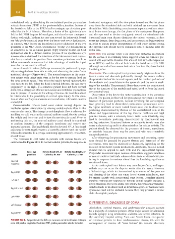
contralateral side by stimulating the contralateral pontine paramedian horizontal nystagmus, with the slow phase toward and the fast phase
reticular formation (PPRF) at the pontomedullary junction. Lesions of away from the stimulated side and with minimal eye movement from
the frontal eye fields or the PPRFs lead to conjugate eye deviation, pro- the midline. With diminishing consciousness in patients without struc-
vided that the MLF is intact. Therefore, a lesion of the right frontal eye tural brain stem damage, the fast phase of the nystagmus disappears,
field or left PPRF impairs leftward gaze, and thus the eyes conjugately and the eyes tend to deviate conjugately toward the stimulated side.
deviate to the right. In short, the eyes turn toward the lesion with frontal Structural brain stem disease eliminates the caloric response, as does
eye field dysfunction and away from the lesion with PPRF dysfunction. inner ear disease, deep drug coma, and anticonvulsant drug overdose.
In contrast, MLF lesions are manifested as poor adduction of the eye In order to ensure proper interpretation of cold water caloric testing,
ipsilateral to the MLF lesion. Spontaneous “roving” eye movements in the opposite side should not be stimulated until 5 minutes after the
all directions in the comatose patient imply bilateral frontal eye field initial side.
dysfunction due to a diffuse cerebral process. If no spontaneous eye Corneal Reflex The corneal reflex is an important protective mechanism
movements are observed, the intactness of the interconnections respon- for the cornea. It is a blinking reflex triggered when the cornea is pre-
sible for eye control is in question. Since comatose patients are unable to sented with any tactile stimulus. The afferent limb is via the trigeminal
follow commands, maneuvers that take advantage of vestibular input nerve (CN V), and the efferent limb is via the facial nerve (CN VII).
to ocular control must be utilized. Although corneal reflexes assess brain stem function, they have limited
An oculocephalic reflex (doll’s eye maneuver) is performed by rap- localizing value.
idly rotating the head from side to side, and observing the patient’s eye
positional changes (Figure 88-3). The normal response in the coma- Motor Function The corticospinal tract predominantly originates from the
tose patient with intact brain stem is for the eyes to remain fixed on frontal cortex and descends ipsilaterally through the corona radiata,
the same point in space. Thus, when the head is turned rightward, the the posterior limb of the internal capsule, and the cerebral peduncle of
eyes move to the left. When the head is turned leftward, the eyes move the midbrain and consolidates in the pyramids, and the ventral swell-
conjugately to the right. If a comatose patient does not have normal ings of the medulla. The pyramidal fibers decussate to the contralateral
doll’s eyes, a disruption of brain stem ocular and vestibular connections side at the junction of the medulla and spinal cord to form the lateral
may be present. Of course, in the setting of trauma, the head should not corticospinal tract.
be rotated due to the possibility of cervical spine injury. In this situa- Observation is the key to the motor examination in the comatose
tion or when doll’s eye maneuvers are inconclusive, cold water calorics patient. The patient is observed for spontaneous movements or main-
are helpful. tenance of particular postures. Lesions involving the corticospinal
Oculovestibular reflexes (cold water caloric testing) depend on tract generally lead to diminished contralateral spontaneous activ-
vestibular system stimulation by altering endolymphatic flow in the ity. Upper midbrain or more rostral lesions may lead to decorticate
semicircular canals. The change in endolymphatic flow is achieved by posturing characterized by flexion of the contralateral arm at the
instilling ice-cold water in the external auditory canal, thereby cooling elbow and hyperextension of the leg. Central midbrain and high
the middle and inner ear, and in turn the semicircular canal. Prior to pontine lesions, with a relatively intact brain stem inferiorly, may
performing this test, the external auditory canal should be examined lead to decerebrate posturing characterized by contralateral arm
to confirm intactness of the tympanic membrane and remove any and leg extension. Structural lesions or metabolic insults may also
impacted cerumen. The head should then be elevated 30°. A functional cause such posturing and it is often mistaken for seizure activity. The
apparatus for instilling the water is a butterfly catheter (with the needle patient should be observed for the presence of tremor, myoclonus,
removed) connected to a syringe containing approximately 15 to 20 mL or asterixis, because these may be associated with toxic-metabolic
of cold water. encephalopathies.
The responses to cold water in patients with various lesions are After observing for spontaneous movements and posturing, motor
summarized in Figure 88-3. In normal wakeful patients, the response is tone should be assessed by passive flexion and extension of the
extremities. Tone may be increased or decreased, depending on the
location of the motor system involvement. Afterward, noxious stimuli
should then be applied to each limb and the supraorbital regions.
Basal eye Rotate head left, or Rotate head right, or Purposeful movement upon noxious stimulation suggests intactness
Lesion position Calorics, Rt. ear Calorics, Lt. ear
of motor tracts to that limb, whereas decorticate or decerebrate pos-
turing in response to noxious stimuli has the localizing significance
Normal mentioned above.
response
Acute corticospinal tract lesions may cause hyporeflexia, and hyper-
reflexia may not occur for days to weeks after the injury. However,
a Babinski sign, which is characterized by extension of the great toe
Right MLF
and fanning of the other toes upon lateral plantar stimulation, may
be present acutely with corticospinal tract lesions. Complete bilateral
Left MLF paralysis without any response to noxious stimuli usually indicates a
grave prognosis. However, spinal cord injury, neuromuscular transmis-
sion blockade, or an illness such as myasthenia gravis or Guillain-Barré
Right frontal
syndrome must not be excluded because they may produce a similar
state of complete paralysis.
Left PPRF
DIFFERENTIAL DIAGNOSIS OF COMA
Left frontal
Alcoholism, cerebral trauma, and cerebrovascular diseases account
for a majority of comatose patients. Other major causes for admission
Right PPRF include epilepsy, drug intoxication, diabetes, and severe infection. In
the university hospital setting Plum and Posner found one-quarter
FIGURE 88-3. Eye positions in the doll’s eye maneuver and with cold caloric testing in of comatose patients to have cerebrovascular disease, 6% were the
coma. MLF, median longitudinal fasciculus; PPRF, pontine paramedian reticular formation. consequence of trauma, all “mass lesions” (ie, tumors, abscesses,
section06.indd 834 1/23/2015 12:56:23 PM