
Page 1195 - Hall et al (2015) Principles of Critical Care-McGraw-Hill
P. 1195
832 PART 6: Neurologic Disorders
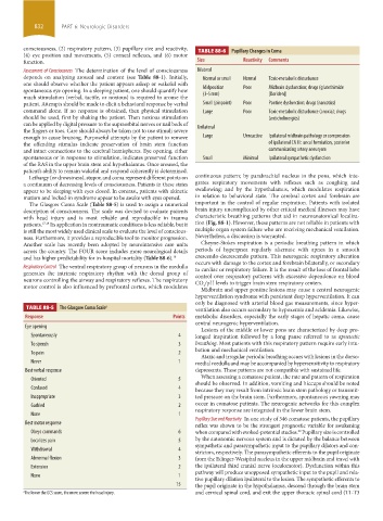
consciousness, (2) respiratory pattern, (3) pupillary size and reactivity, TABLE 88-6 Pupillary Changes in Coma
(4) eye position and movements, (5) corneal reflexes, and (6) motor
function. Size Reactivity Comments
Assessment of Consciousness The determination of the level of consciousness Bilateral
depends on analyzing arousal and content (see Table 88-1). Initially, Normal or small Normal Toxic-metabolic disturbance
one should observe whether the patient appears asleep or wakeful with
spontaneous eye opening. In a sleeping patient, one should quantify how Midposition Poor Midbrain dysfunction; drugs (glutethimide
much stimulation (verbal, tactile, or noxious) is required to arouse the (3-5 mm) [Doriden])
patient. Attempts should be made to elicit a behavioral response by verbal Small (pinpoint) Poor Pontine dysfunction; drugs (narcotics)
command alone. If no response is obtained, then physical stimulation Large Poor Toxic-metabolic disturbance (anoxia); drugs
should be used, first by shaking the patient. Then noxious stimulation ( anticholinergics)
can be applied by digital pressure to the supraorbital nerves or nail beds of Unilateral
the fingers or toes. Care should always be taken not to use stimuli severe
enough to cause bruising. Purposeful attempts by the patient to remove Large Unreactive Ipsilateral midbrain pathology or compression
the offending stimulus indicate preservation of brain stem function of ipsilateral CN III: uncal herniation, posterior
and intact connections to the cerebral hemispheres. Eye opening, either communicating artery aneurysm
spontaneous or in response to stimulation, indicates preserved function Small Minimal Ipsilateral sympathetic dysfunction
of the RAS in the upper brain stem and hypothalamus. Once aroused, the
patient’s ability to remain wakeful and respond coherently is determined.
Lethargy (or drowsiness), stupor, and coma represent different points on continuous pattern; by parabrachial nucleus in the pons, which inte-
a continuum of decreasing levels of consciousness. Patients in these states grates respiratory movements with reflexes such as coughing and
appear to be sleeping with eyes closed. In contrast, patients with akinetic swallowing; and by the hypothalamus, which modulates respiration
mutism and locked-in syndrome appear to be awake with eyes opened. in relation to behavioral state. The cerebral cortex and forebrain are
The Glasgow Coma Scale (Table 88-5) is used to assign a numerical important in the control of regular respiration. Patients with isolated
description of consciousness. The scale was devised to evaluate patients brain injury uncomplicated by other critical medical illnesses may have
with head injury and is most reliable and reproducible in trauma characteristic breathing patterns that aid in neuroanatomical localiza-
patients. 17,18 Its application in nontraumatic conditions is less reliable, but it tion (Fig. 88-1). However, these patterns are not reliable in patients with
is still the most widely used clinical scale to evaluate the level of conscious- multiple organ system failure who are receiving mechanical ventilation.
ness. Furthermore, it provides a reproducible tool to monitor progression. Nevertheless, a discussion is warranted.
Another scale has recently been adopted by neurointensive care units Cheyne-Stokes respiration is a periodic breathing pattern in which
across the country. The FOUR score includes more neurological details periods of hyperpnea regularly alternate with apnea in a smooth
and has higher predictability for in-hospital mortality (Table 88-6). 19 crescendo-decrescendo pattern. This neurogenic respiratory alteration
Respiratory Control The ventral respiratory group of neurons in the medulla occurs with damage to the cortex and forebrain bilaterally, or secondary
to cardiac or respiratory failure. It is the result of the loss of frontal lobe
generates the intrinsic respiratory rhythm with the dorsal group of control over respiratory patterns with excessive dependence on blood
neurons controlling the airway and respiratory reflexes. The respiratory CO /pH levels to trigger brain stem respiratory centers.
motor control is also influenced by prefrontal cortex, which modulates Midbrain and upper pontine lesions may cause a central neurogenic
2
hyperventilation syndrome with persistent deep hyperventilation. It can
only be diagnosed with arterial blood gas measurements, since hyper-
TABLE 88-5 The Glasgow Coma Scale a ventilation also occurs secondary to hypoxemia and acidemia. Likewise,
Response Points metabolic disorders, especially the early stages of hepatic coma, cause
central neurogenic hyperventilation.
Eye opening
Lesions of the middle or lower pons are characterized by deep pro-
Spontaneously 4 longed inspiration followed by a long pause referred to as apneustic
To speech 3 breathing. Most patients with this respiratory pattern require early intu-
bation and mechanical ventilation.
To pain 2
Ataxic and irregular periodic breathing occurs with lesions in the dorso-
Never 1 medial medulla and may be accompanied by hypersensitivity to respiratory
Best verbal response depressants. These patterns are not compatible with sustained life.
When assessing a comatose patient, the rate and pattern of respiration
Oriented 5
should be observed. In addition, vomiting and hiccups should be noted
Confused 4 because they may result from intrinsic brain stem pathology or transmit-
Inappropriate 3 ted pressure on the brain stem. Furthermore, spontaneous yawning may
Garbled 2 occur in comatose patients. The neurogenic networks for this complex
respiratory response are integrated in the lower brain stem.
None 1
Pupillary Size and Reactivity In one study of 346 comatose patients, the pupillary
Best motor response reflex was shown to be the strongest prognostic variable for awakening
Obeys commands 6 when compared with evoked-potential studies. Pupillary size is controlled
20
Localizes pain 5 by the autonomic nervous system and is dictated by the balance between
sympathetic and parasympathetic input to the pupillary dilators and con-
Withdrawal 4
strictors, respectively. The parasympathetic efferents to the pupil originate
Abnormal flexion 3 from the Edinger-Westphal nucleus in the upper midbrain and travel with
Extension 2 the ipsilateral third cranial nerve (oculomotor). Dysfunction within this
pathway will produce unopposed sympathetic input to the pupil and rela-
None 1
tive pupillary dilation ipsilateral to the lesion. The sympathetic efferents to
15 the pupil originate in the hypothalamus, descend through the brain stem
a The lower the GCS score, the more severe the head injury. and cervical spinal cord, and exit the upper thoracic spinal cord (T1-T3
section06.indd 832 1/23/2015 12:56:21 PM